
|
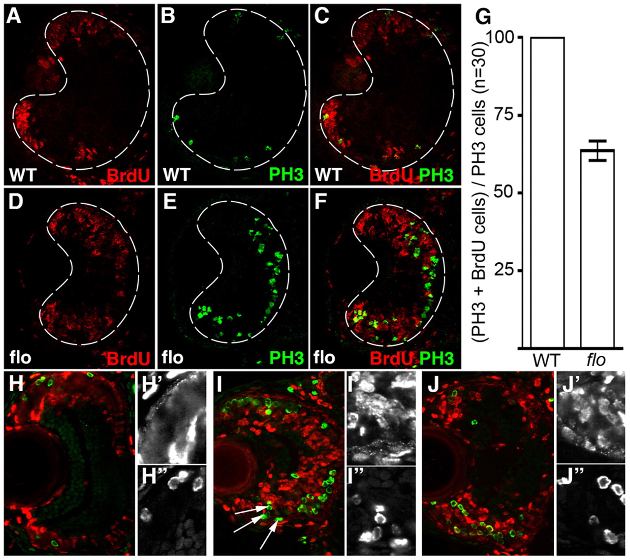
Fig. 5 flo retinal cells fail to progress efficiently through the cell cycle. (A-F,H-J″) Frontal transverse cryosections of zebrafish retina stained for markers of DNA replication (BrdU, red) and M phase (PH3, green). Wild-type (A-C) and flo + p53MO (D-F) embryos were injected with BrdU at 53 hpf and again at 55 hpf, then fixed <75 minutes later. Single-channel magnifications of BrdU (H′,I′,J′) and PH3 (H″,I″,J″) in the CMZ are shown. (G) The average number of PH3+ cells that are also BrdU+ per section (n=30 eyes) was calculated and graphed with error bars (99% confidence limits). All of the PH3+ cells in wild-type siblings were also BrdU+. Significantly fewer cells (27.5%) were double labelled in flo embryos (P<0.0001, Student′s t-test). As above, wild-type (H), flo (I) and flo + p53MO (J) embryos were injected with BrdU but then fixed 24 hours later.