
|
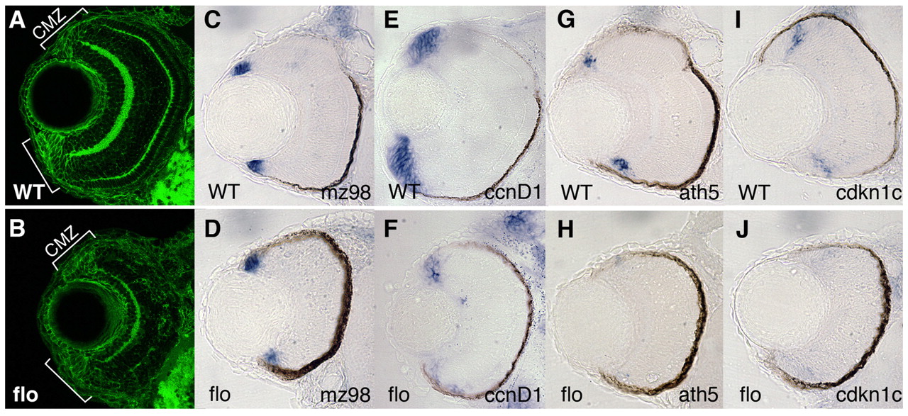
Fig. 3 CMZ cells in flo embryos show reduced expression of cycling and differentiation markers. (A,B) CMZ cells are present in flo retinas. Frontal transverse cryosections of zebrafish retinae stained for β-catenin (green). Brackets denote CMZ boundaries. (C-J) Gene expression in subpopulations of cells within the CMZ. mz98, a marker of the peripheral, putative stem cell compartment, is expressed in wild-type siblings (C) and flo (D) embryos. ccnd1 is highly expressed in the wild-type proliferating progenitors of the CMZ (E) but is reduced in flo eyes (F). ath5 (G,H) and cdkn1c (I,J) are expressed in cells in central CMZ concomitant with the final division cycle in the wild-type CMZ (G,I), but are absent in the flo retina (H,J).