
|
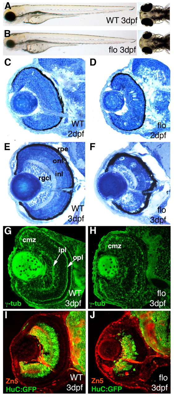
Fig. 1 flo mutants have small eyes. (A,B) Lateral (left) and dorsal (right) views of live wild-type (A) and flo (B) zebrafish embryos at 3 dpf. (C-F) Frontal transverse sections through wild-type (C,E) and flo (D,F) embryos at 2 dpf (C,D) and 3 dpf (E,F). (G-J) Frontal cryosections of wild-type (G,I) and flo (H,J) eyes immunostained to detect markers for polarity (G,H, γ-tubulin, green) and differentiation [I,J; zn5 (Alcama), RGCs, red; HuC::GFP (Elavl3), RGCs and amacrine cells, green]. rgcl, retinal ganglion cell layer; inl, inner nuclear layer; onl, outer nuclear layer; rpe, retinal pigmented epithelium; cmz, ciliary marginal zone; ipl, inner plexiform layer; opl, outer plexiform layer.