
|
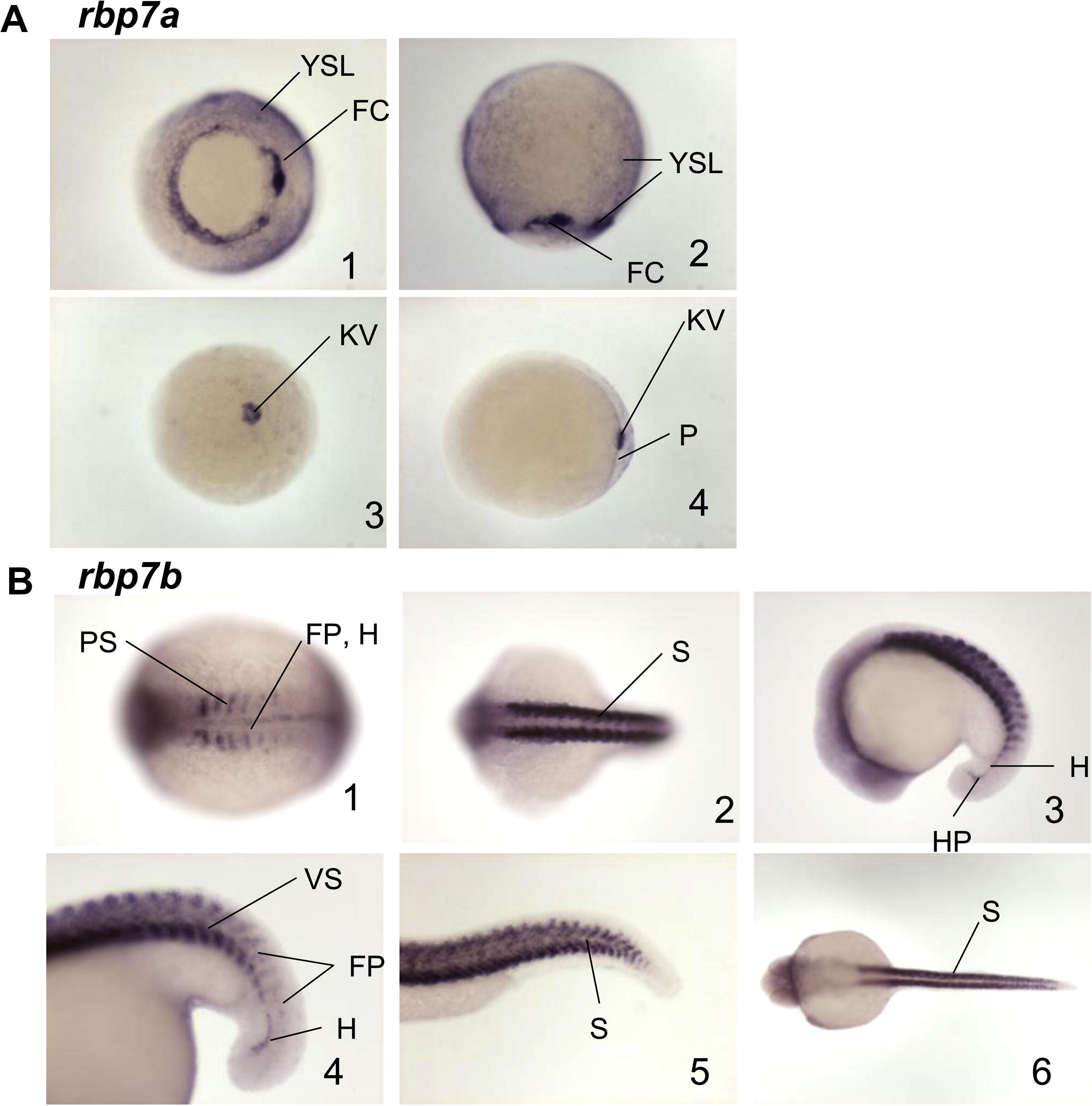
Fig. 6 Spatio-temporal distribution of rbp7a and rbp7b gene transcripts in zebrafish embryos as determined by whole mount in situ hybridization. rbp7a transcripts were seen in the yolk syncytial layer (YSL) and forerunner cells (FC) at the gastrula stage at 4 hpf, ventral (A1) and lateral (A2) planes. At 12 hpf, rbp7a transcripts were detected in Kupffer’s vesicle (kV) and the periderm (P), dorsal (A3) and lateral (A4) views. rbp7b was first detected 10 hpf in early somitogenesis, in the floor plate, (FP), hypochord (H), and posterior part of the somites (PS), dorsal plane (B1). In middle somitogenesis, rbp7b transcript was detected in somites (S), hypochord, hypochord precursors (HP), and ventral part of somites (VS), dorsal (B2) and lateral (B3 and B4) planes. At 24 hpf, rbp7b transcripts were detected in the somites, lateral (B5) and dorsal (B6) planes.
Reprinted from Gene expression patterns : GEP, 10(4-5), Belliveau, D.J., Venkatachalam, A.B., Thisse, C., Thisse, B., Ma, H., and Wright, J.M., The duplicated retinol-binding protein 7 (rbp7) genes are differentially transcribed in embryos and adult zebrafish (Danio rerio), 167-176, Copyright (2010) with permission from Elsevier. Full text @ Gene Expr. Patterns