
Fig. 4
- ID
- ZDB-IMAGE-100616-114
- Genes
- Publication
- Petzold et al., 2010 - SCORE imaging: specimen in a corrected optical rotational enclosure
- All Figures
- Figures for Petzold et al., 2010
|
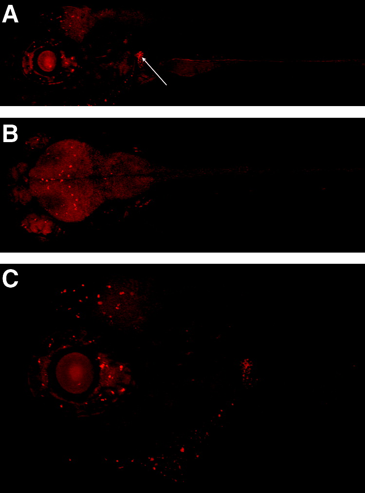
Fig. 4 SCORE imaging of fluorescent, living zebrafish. Imaging SEC0124 of a 5 dpf mRFP-labeled protein trap zebrafish housed in FEP tubing. (A) Sagittal image of an mRFP-labeled protein trap zebrafish shows enhancement in specific locations in the neural region as well as a bright kidney (Arrow). (B) Coronal image of an mRFP-labeled protein trap zebrafish includes ubiquitous neural expression with enhancement in specific locations. (C) Sagittal image of an mRFP-labeled protein trap zebrafish shows a general neural haze with enhancement in specific locations at 100x magnification (also see Supplemental Movie S5 [showing the spatial location of RFP patterning], available online at www.liebertonline.com).