
IMAGE
Fig. 9
- ID
- ZDB-IMAGE-100527-11
- Antibodies
- Publication
- Chao et al., 2010 - A male with unilateral microphthalmia reveals a role for TMX3 in eye development
- All Figures
- Figures for Chao et al., 2010
Image
|
Figure Caption
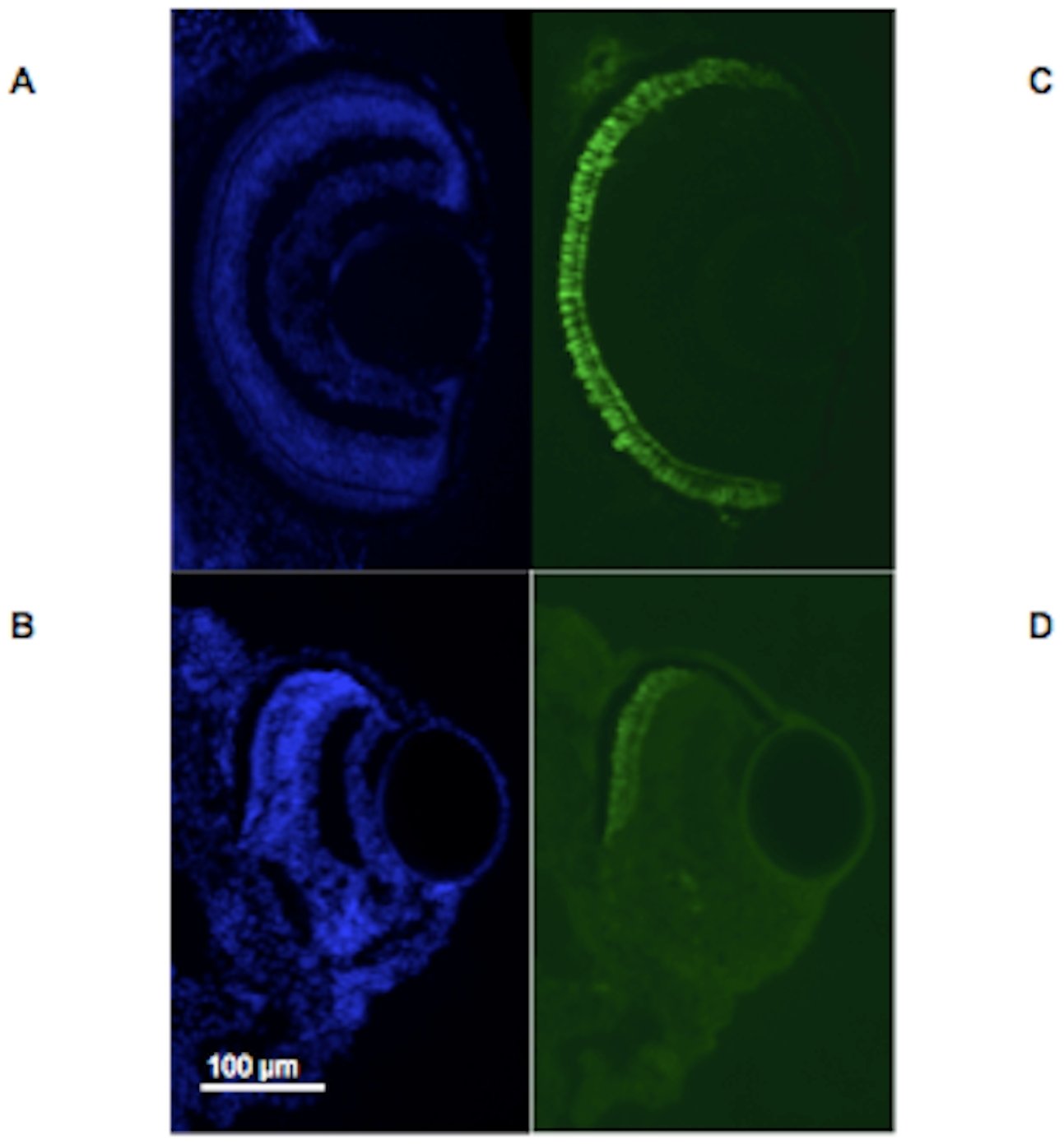
Fig. 9 Morphant larvae show altered formation of the ventral eye.
Fig. 9A–D. Fig. 9A–B. Staining of control injected zebrafish eye with DAPI and FITC at 6 dpf to image zpr-1 shows a strong signal that can be seen at the ciliary margins of the retina. Fig. 9C–D. Staining of anti-ATG morphant (MO1) zebrafish eye with DAPI and FITC at 6 dpf to image zpr-1 shows absent signal for zpr-1 at the ventral region of the retina in an eye with a coloboma, whereas staining at the dorsal region of the retina appears normal. Fish are oriented so that the ventral surface of the eye is seen inferiorly in each photograph.
Figure Data
Acknowledgments
This image is the copyrighted work of the attributed author or publisher, and
ZFIN has permission only to display this image to its users.
Additional permissions should be obtained from the applicable author or publisher of the image.
Full text @ PLoS One