
Fig. S1
- ID
- ZDB-IMAGE-100506-9
- Publication
- Nguyen et al., 2010 - The Paf1 complex component Leo1 is essential for cardiac and neural crest development in zebrafish
- All Figures
- Figures for Nguyen et al., 2010
|
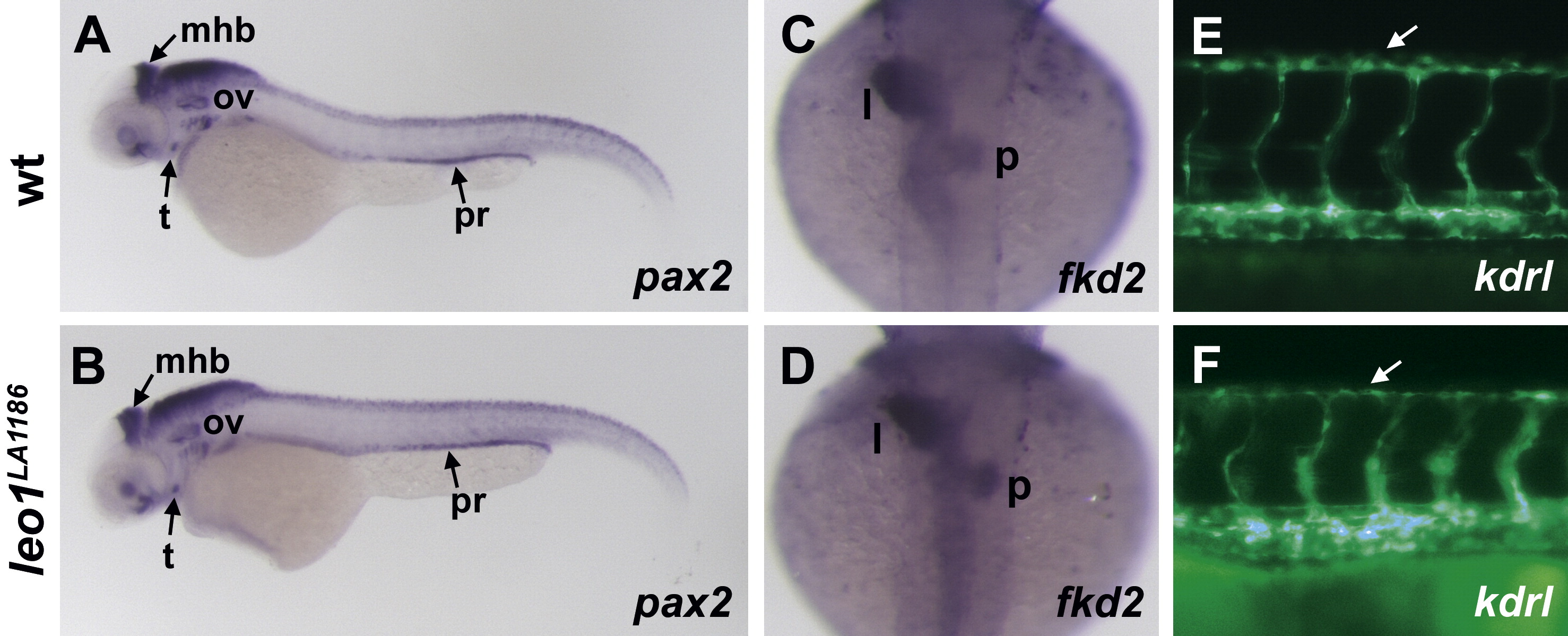
Fig. S1 Leo1LA1186 mutants do not have general developmental delay. (A, B) pax2 expression is detected in the midbrain–hindbrain boundary (mhb), thyroid (t), otic vesicle (ov), and pronephric ducts (pr) of wild-type embryos (A) at 50 hpf. Leo1LA1186 mutants have normal expression of pax2 in all of these structures at 50 hpf (B), indicating that the timely organogenesis of the brain, thyroid, otic vesicle, and pronephric ducts does not require Leo1 activity. (C, D) At 50 hpf, both wild-type (C) and leo1LA1186 mutant (D) embryos have a leftward-looped gut, liver (l), and pancreas (p) labeled by fkd2 staining, demonstrating that the morphogenesis of the gut is not developmentally delayed upon loss of Leo1 activity. (E, F) By 50 hpf, the primary intersegmental vessels have branched dorsally to form the dorsal longitudinal anastomotic vessel (arrows) in both wild-type (E) and leo1LA1186 mutant (F) embryos, suggesting that Leo1 activity is not essential for patterning of the trunk vasculature within a normal developmental timeframe. Vessels in E and F are visualized by fluorescence microscopy of Tg(kdrl:GFP)la116 transgenic embryos.
Reprinted from Developmental Biology, 341(1), Nguyen, C.T., Langenbacher, A., Hsieh, M., and Chen, J.N., The Paf1 complex component Leo1 is essential for cardiac and neural crest development in zebrafish, 167-175, Copyright (2010) with permission from Elsevier. Full text @ Dev. Biol.