
Fig. S2
- ID
- ZDB-IMAGE-100506-38
- Publication
- Nakano et al., 2010 - Biogenesis of GPI-anchored proteins is essential for surface expression of sodium channels in zebrafish Rohon-Beard neurons to respond to mechanosensory stimulation
- All Figures
- Figures for Nakano et al., 2010
|
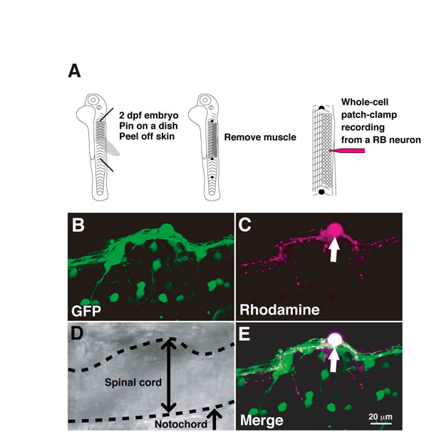
Fig. S2 Whole-cell patch-clamp recording from RB neurons. (A) Schematic summary of the experimental procedures. Embryos (2 dpf) were pinned on a silicone dish and their skin and muscle were removed. A glass electrode was applied to a GFP-positive RB neuron to record from a single RB cell. (B-E) The patched neurons were identifiable by their morphology. GFP-expressing neurons with large cell bodies located at the dorsal spinal cord were used for whole-cell patch-clamp recording. After whole-cell recording, each patched cell was morphologically identified as an RB neuron. (B) GFP fluorescence. (C) Rhodamine fluorescence diffused from a patch electrode. (D) DIC image. (E) A merged image of GFP and rhodamine confirmed that the patched cell is an RB neuron. Arrows represent patch-clamped RB cells.