
Fig. S1
- ID
- ZDB-IMAGE-100506-37
- Publication
- Nakano et al., 2010 - Biogenesis of GPI-anchored proteins is essential for surface expression of sodium channels in zebrafish Rohon-Beard neurons to respond to mechanosensory stimulation
- All Figures
- Figures for Nakano et al., 2010
|
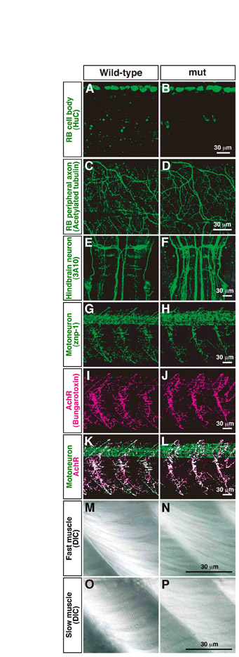
Fig. S1 Morphology of RB neurons, CNS neurons and muscle is normal in mi310 mutants. (A-D) Anti-HuC and anti-acetylated tubulin labeled cell bodies and peripheral axons of RB neurons, respectively, in both wild-type (A,C) and mutant (B,D) embryos. (E,F) Reticulospinal neurons in hindbrain were visualized with 3A10 antibody. (G-L) Motoneurons and nicotinic acetylcholine receptors were labeled with znp-1 (G,H) and α-bungarotoxin (I,J), respectively, and their colocalizations represented neuromuscular junctions (K,L). (M-P) Fast muscle fibers (M,N) and slow muscle fibers (O,P) were visualized by DIC in both wild-type and mutant muscle.