
Fig. 5
- ID
- ZDB-IMAGE-100506-29
- Genes
- Publication
- Faucherre et al., 2010 - Multispectral four-dimensional imaging reveals that evoked activity modulates peripheral arborization and the selection of plane-polarized targets by sensory neurons
- All Figures
- Figures for Faucherre et al., 2010
|
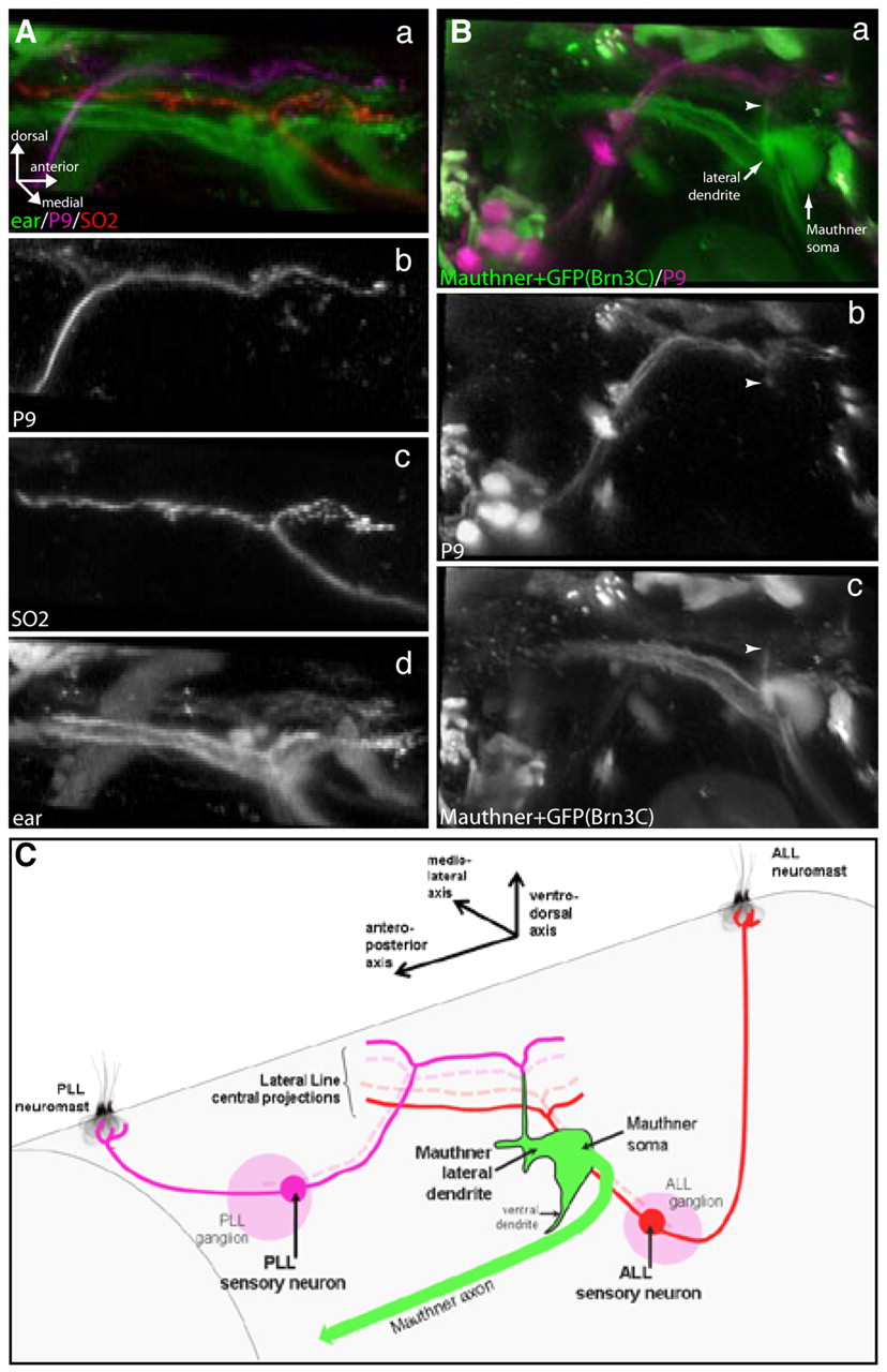
Fig. 5 Central projections of first-order lateral line sensory neurons in tmie mutants. Central somatotopy and targeting of the Mauthner cell are conserved in tmie mutants. (Aa-d) Fluorescent dextran injections in tmie-/- brn3c:gfp transgenic fish showing somatotopy of first-order lateral line sensory neuron projections coming from posterior (b) and anterior (c) neuromasts. Projections coming from P9 (PLL) neuromasts (b; Alexa 647-dextran) and SO2 (ALL) neuromasts (c; Rhodamine-dextran) project in a dorsoventral manner (a) in the hindbrain. (d) Projections coming from the ear (Brn3c:GFP). (Ba-c) Fluorescent dextran injections in tmie-/- brn3c:gfp transgenic fish showing central targeting of the Mauthner lateral dendrite by posterior and anterior lateral line projections (a). Projections coming from the P9 (PLL) neuromast (b; Alexa 647-dextran) connect (arrowheads) to the lateral-most portion of the Mauthner cell lateral dendrite (c; fluorescein-dextran), which extends ventrodorsally. (C) Scheme (not to scale) depicting the central somatotopy of the first-order lateral line sensory neuron projections, and their targeting of the Mauthner cell lateral dendrite. In tmie fish, the anteroposterior axis of the lateral line neuromast projects onto the ventrodorsal axis of the Mauthner lateral dendrite dorsal projection, as in wild-type fish. ALL, anterior lateral line; PLL, posterior lateral line.