
Fig. 2
- ID
- ZDB-IMAGE-100506-28
- Genes
- Publication
- Faucherre et al., 2010 - Multispectral four-dimensional imaging reveals that evoked activity modulates peripheral arborization and the selection of plane-polarized targets by sensory neurons
- All Figures
- Figures for Faucherre et al., 2010
|
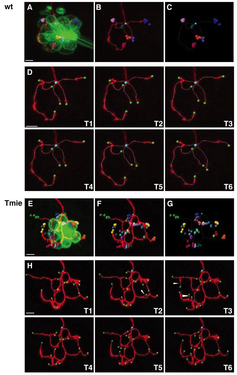
Fig. 2 Four-dimensional reconstruction of a single labeled afferent neuron. (A-G) Four-dimensional representations of an afferent neuron in wild-type (A-C) and tmie mutant (E-G) fish. (A,E) mem-TdTomato-labeled neuron contacting the GFP-expressing hair cells. The terminal point of each neuron has been plotted with a different color. For each terminal point (i.e. for each color) positions have been plotted over time, every 10 minutes for 1 hour. (B,F) Same as A,E but without the hair cells. (C,G) Same as B,F but with only the dots representing the terminal point. The unique cyan dot indicates the beginning point of the neuronal arbor. The white frame corresponds to the neuronal arbor reconstruction of the first time frame. (D,H) Maximal projections and neuronal arbor reconstruction of the same neuron over time. Images T1-T6 were taken every 10 minutes for 1 hour. Beginning point is in cyan, terminal points are in green, branching points are in red, and special branching points (multiple bifurcations) are in blue. Arrowheads indicate examples of transient neurites. Scale bars: 5 μm.