
|
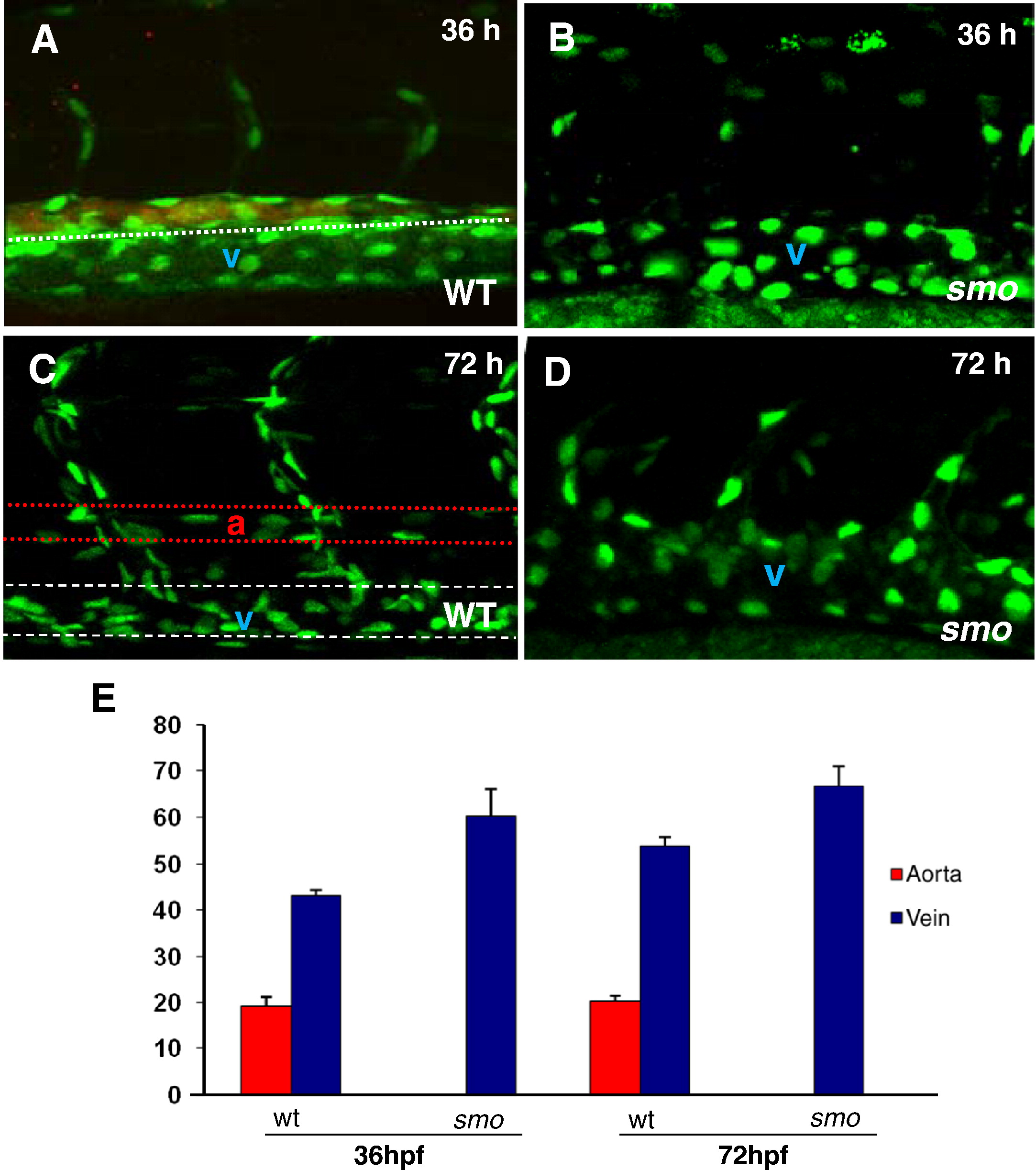
Fig. 3 The increase of venous cells is equivalent to loss of arterial cells in smo mutants. (A–D) Confocal optics displaying individual nuclei of endothelial cells in the dorsal aorta (A, B stained with efnb2 antibody, the posterior cardinal vein and intersomitic vessels in wild-type embryos [Tg(fli:EGFP-nuc)] at 36 hpf (A) and 72 hpf (C),as compared to the increased endothelial nuclei in enlarged cardinal veins in smo mutants [smohi1640/smohi1640;Tg(fli:EGFP-nuc)] at 36 hpf (B) and 72 hpf (D). (E) Bar chart depicting number of arterial and venous cells in wild-type embryos compared to smo mutants. At 36 hpf, arterial cell number: 19.2 ± 1.7 (WT), 0 (smo); Venous cell number: 43.2 ± 1.3 (WT), 60.3 ± 2.4 (smo). At 72 hpf, arterial cell number: 20.2 ± 1.3 (WT), 0 (smo); Venous cell number: 54 ± 1.6 (WT), 66.8 ± 3.4 (smo). Endothelial cells were counted from 12 lateral sections derived from 6 wild-type embryos and 6 smo mutants. Each section covers four segment lengths of intersomitic vessels along the dorsal aorta and the posterior cardinal vein in the middle trunk above the yolk extension region. a: aorta. v: vein.
Reprinted from Developmental Biology, 341(1), Williams, C., Kim, S.H., Ni, T.T., Mitchell, L., Ro, H., Penn, J.S., Baldwin, S.H., Solnica-Krezel, L., and Zhong, T.P., Hedgehog signaling induces arterial endothelial cell formation by repressing venous cell fate, 196-204, Copyright (2010) with permission from Elsevier. Full text @ Dev. Biol.