
Fig. 3
- ID
- ZDB-IMAGE-100504-3
- Publication
- Tay et al., 2010 - A vertebrate-specific Chp-PAK-PIX pathway maintains E-cadherin at adherens junctions during zebrafish epiboly
- All Figures
- Figures for Tay et al., 2010
|
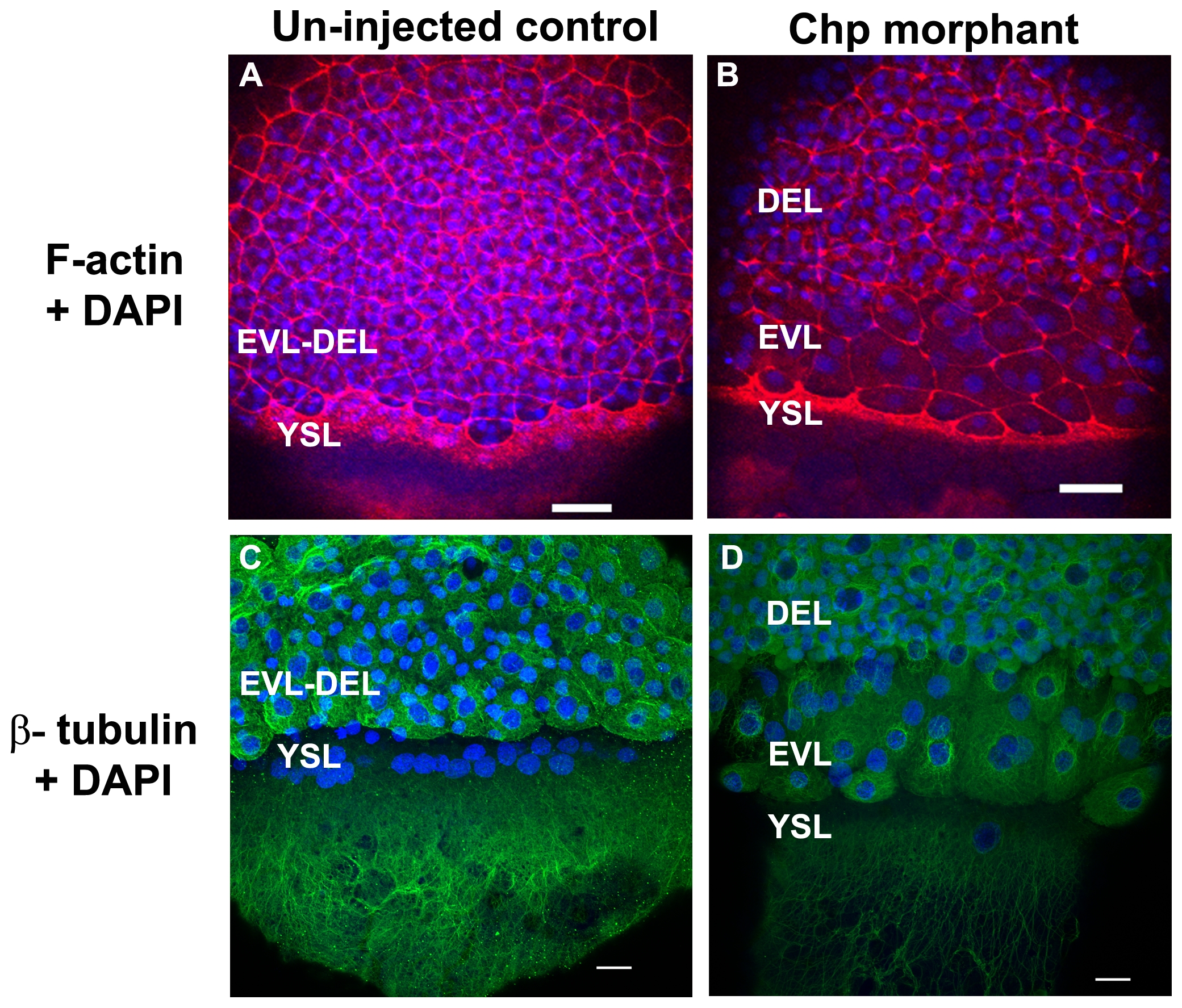
Fig. 3 The actin cytoskeleton and microtubule networks in Chp morphants.
Low resolution confocal images (10x objective) comparing (A) un-injected control and (B) Chp morphant with respect to F-actin organization at the vegetal margins of EVL and DEL, and at the external yolk syncytial layer (YSL). The deep cells marked by DAPI stained nuclei, fail to properly migrate in Chp morphants (B and D). As a result the cell margin is thinner and the F-actin ring of the EVL and YSL appears more compact. Confocal images at higher magnification (40x) comparing (C) un-injected control and (D) Chp morphant stained with β-tubulin. Microtubule organization in Chp morphant was largely normal. Scale bars represent 40 μm in panels A-B and 20 μm in panels C-D.