
|
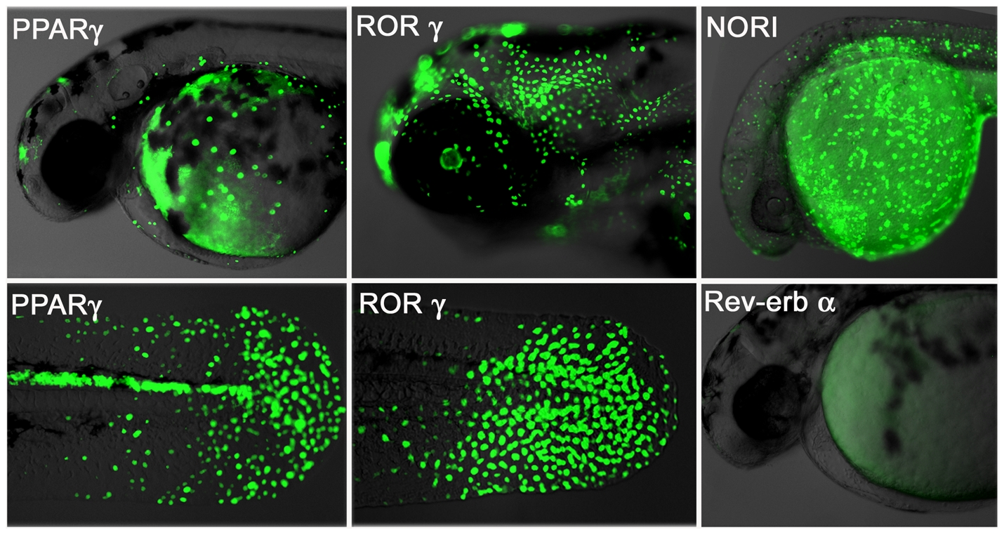
Fig. 2 Human NRs interact with fish ligands and cofactors.
Transgenic NR zebrafish show unique GFP patterns at different stages of development. Activity patterns of the PPARγ (first panel), RORγ (middle panel), NOR1 (upper right picture) and Rev-erbα (lower right picture) LT constructs. Embryos were heat pulsed at 37°C for 30 min and images taken 24 h later. PPARγ (48 hpf, F2) embryos show GFP expression in cells of the epidermis and heart, as well as in the posterior spinal cord. Strong GFP expression occurs in the epidermis and retina of RORγ embryos (72 hpf, F2). NOR1 embryos (F3) express GFP in the posterior spinal cord, hatching gland, epidermis and yolk syncictial layer (24 hpf, F2). Rev-erbα embryos show no GFP expression. Overlay pictures of bright field and GFP (75% transparent) are shown. Views are lateral with anterior to the left.