
Fig. 3
- ID
- ZDB-IMAGE-100429-50
- Genes
- Publication
- Wada et al., 2010 - Dermal morphogenesis controls lateral line patterning during postembryonic development of teleost fish
- All Figures
- Figures for Wada et al., 2010
|
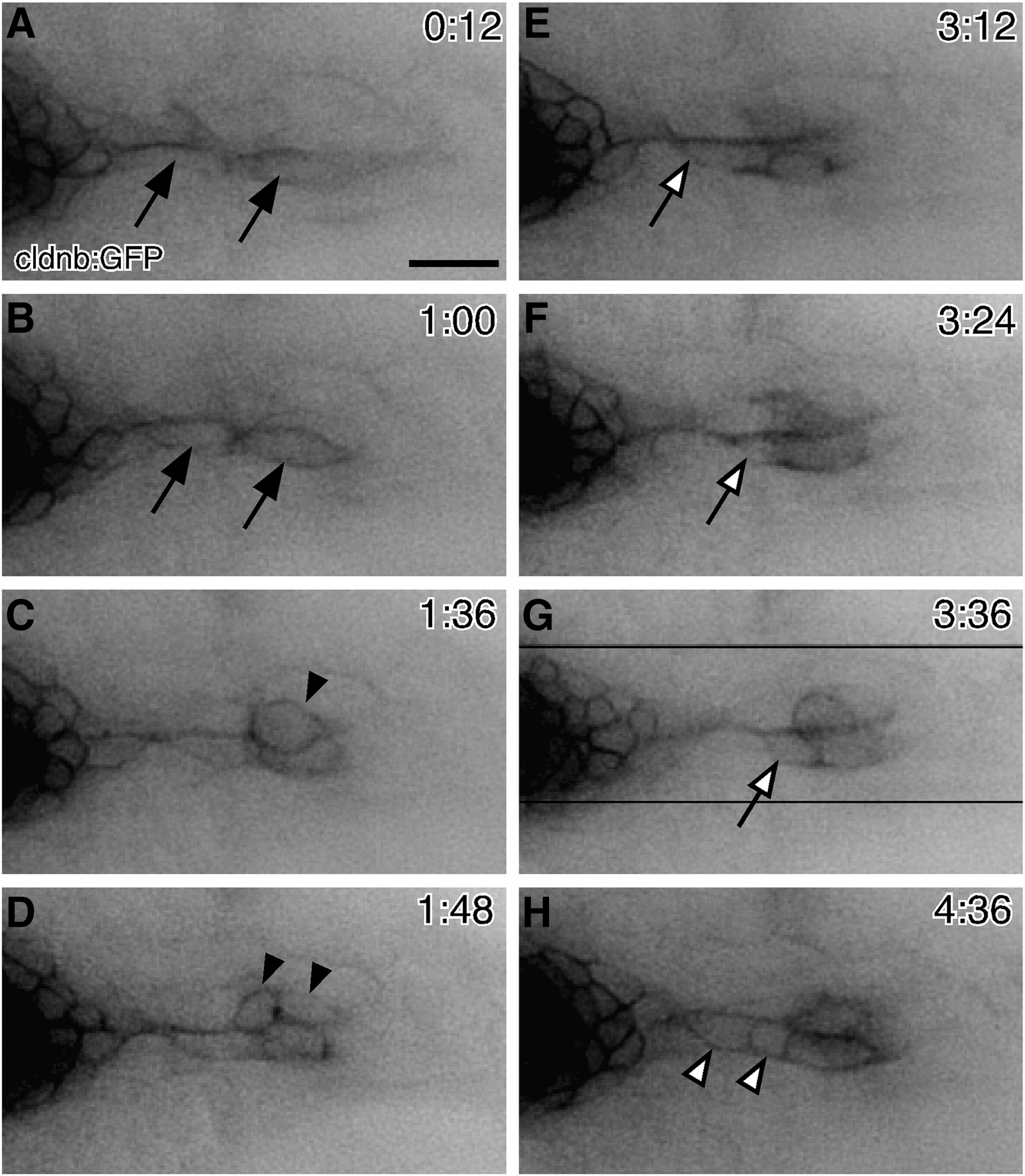
Fig. 3 Behaviors of neuromast cells during a budding process. (A–H) Time-lapse observation of 3-dpf cldnb:gfp transgenic embryo. Images were selected from the original movie (Supplemental Movie S1). (A, B) Two elongated cells are extending from OP1 (indicated by arrows). (C, D) One of these cells becomes round shaped (C, arrowhead) and undergoes a cell division at a distal site (D, arrowheads). (E–G) A cell migrating distally along the elongated cells is indicated by open arrows. (H) Subsequently, cells are recruited proximally (open arrowheads). Time after the start of recording is indicated in each panel. The direction of budding is shown to the right. Scale bar: 20 μm.
Reprinted from Developmental Biology, 340(2), Wada, H., Ghysen, A., Satou, C., Higashijima, S.I., Kawakami, K., Hamaguchi, S., and Sakaizumi, M., Dermal morphogenesis controls lateral line patterning during postembryonic development of teleost fish, 583-594, Copyright (2010) with permission from Elsevier. Full text @ Dev. Biol.