
Fig. S6
- ID
- ZDB-IMAGE-100429-47
- Publication
- Pillay et al., 2010 - The Hox cofactors Meis1 and Pbx act upstream of gata1 to regulate primitive hematopoiesis
- All Figures
- Figures for Pillay et al., 2010
|
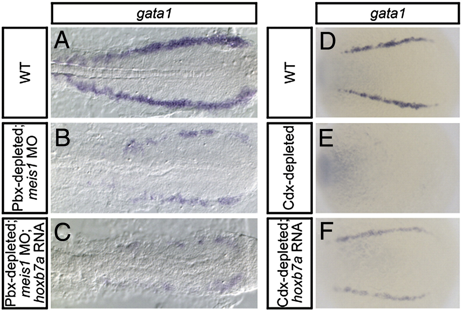
Fig. S6 Overexpressing hoxb7a rescues gata1 erythroid gene expression in Cdx-depleted embryos, but not in Pbx-depleted; meis1-morphant embryos. Shown are representative embryos following in situ hybridization analysis of gata1 expression. (A–C) Dorsal view of PLM gene expression is shown in 16 hpf flat-mounted and deyolked embryos, with anterior oriented to the left. Both Pbx-depleted; meis1-morphant embryos (B) and hoxb7a RNA-injected Pbx-depleted; meis1-morphant embryos (C) exhibit a severe decrease in gata1 expression when compared to wild type embryos (WT; A). (D–F) Dorsal view of PLM gene expression is shown 12 hpf whole-mount embryos, with anterior oriented to the left. Cdx-depleted embryos (E) exhibit a notable decrease in gata1 expression when compared to WT embryos (D). Cdx-depleted, hoxb7a RNA-injected embryos (F) exhibit greater levels of gata1 expression than Cdx-depleted embryos (E).
Reprinted from Developmental Biology, 340(2), Pillay, L.M., Forrester, A.M., Erickson, T., Berman, J.N., and Waskiewicz, A.J., The Hox cofactors Meis1 and Pbx act upstream of gata1 to regulate primitive hematopoiesis, 306-317, Copyright (2010) with permission from Elsevier. Full text @ Dev. Biol.