
Fig. 2
- ID
- ZDB-IMAGE-100429-35
- Publication
- Pillay et al., 2010 - The Hox cofactors Meis1 and Pbx act upstream of gata1 to regulate primitive hematopoiesis
- All Figures
- Figures for Pillay et al., 2010
|
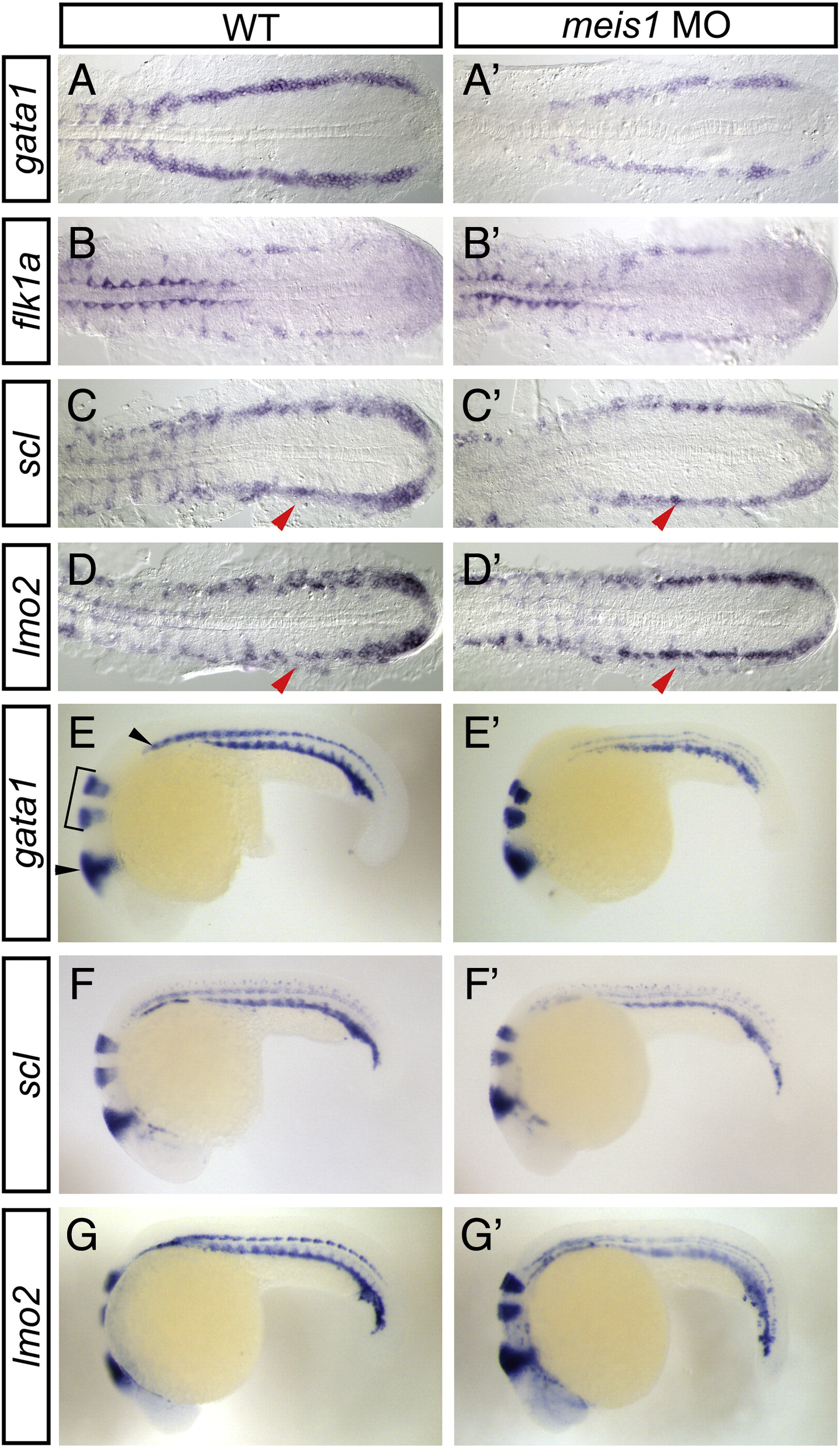
Fig. 2 meis1-morphant embryos exhibit defects in primitive hematopoietic gene expression. Shown are representative embryos following in situ hybridization analysis of hematopoietic marker expression in wild type (WT; A–G) compared with meis1-morphant (A′–G′) embryos. (A–D′) The PLM of 16 hpf flat-mounted, deyolked embryos is shown in dorsal view with anterior to the left. gata1 expression is severely reduced in meis1-morphant embryos (A′; 85%, n = 39) when compared to WT (A). flk1a expression is unchanged in meis1-morphant embryos (B′; 100%, n = 21) when compared to WT (B). Lateral domain of scl (90%, n = 29) and lmo2 expression (79%, n = 24) is abolished in meis1-morphant embryos (C′, D′; red arrowheads), while the medial domain of scl and lmo2 expression is near normal. (E–G′) 24 hpf whole-mount embryos are shown in lateral view with anterior to the left. eng2a expression in the midbrain hindbrain boundary and muscle pioneers (E; black arrowheads) and egr2b expression in hindbrain rhombomeres 3 and 5 (E; bracket) is shown in all panels. gata1 (E′; 91%, n = 53), scl (F′; 97%, n = 35), and lmo2 (G′; 92%, n = 36) expression is reduced in the ICM of meis1-morphant embryos when compared to WT (E–G).
Reprinted from Developmental Biology, 340(2), Pillay, L.M., Forrester, A.M., Erickson, T., Berman, J.N., and Waskiewicz, A.J., The Hox cofactors Meis1 and Pbx act upstream of gata1 to regulate primitive hematopoiesis, 306-317, Copyright (2010) with permission from Elsevier. Full text @ Dev. Biol.