
Fig. S1
- ID
- ZDB-IMAGE-100422-68
- Publication
- Lamont et al., 2010 - Hedgehog signaling via angiopoietin1 is required for developmental vascular stability
- All Figures
- Figures for Lamont et al., 2010
|
Fig. S1
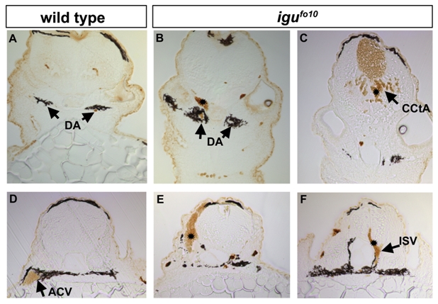
Multiple vessel origins for hemorrhages in igufo10 mutants at 48- 52 hpf
Cross sections of Isolectin B4 staining of blood in the hind brain in the vicinity of the ear (A-C), or in the posterior hindbrain/ anterior trunk (D-F) show hemorrhages in diverse tissue locations of igu mutants. (A-B) Hemorrhages are seen dorsal to the dorsal aorta (DA), but exterior to the brain in (B), as compared to blood confined to the dorsal aorta in wild-type (A). (C) Hemorrhages are also occasionally seen in the vicinity of the cerebellar central arteries (CCtA) within the brain, leading to accumulation of blood in the hindbrain ventricle. (D) More posteriorly, robust circulation can be observed in the anterior cardinal vein of a wild type embryo. (E) A hemorrhage in the somitic tissue is observed in one igu mutant, (F), while a hemorrhage deriving from an intersomitic vessel (ISV) of the trunk is also seen.
Reprinted from Mechanisms of Development, 127(3-4), Lamont, R.E., Vu, W., Carter, A.D., Serluca, F.C., MacRae, C.A., and Childs, S.J., Hedgehog signaling via angiopoietin1 is required for developmental vascular stability, 159-168, Copyright (2010) with permission from Elsevier. Full text @ Mech. Dev.