
|
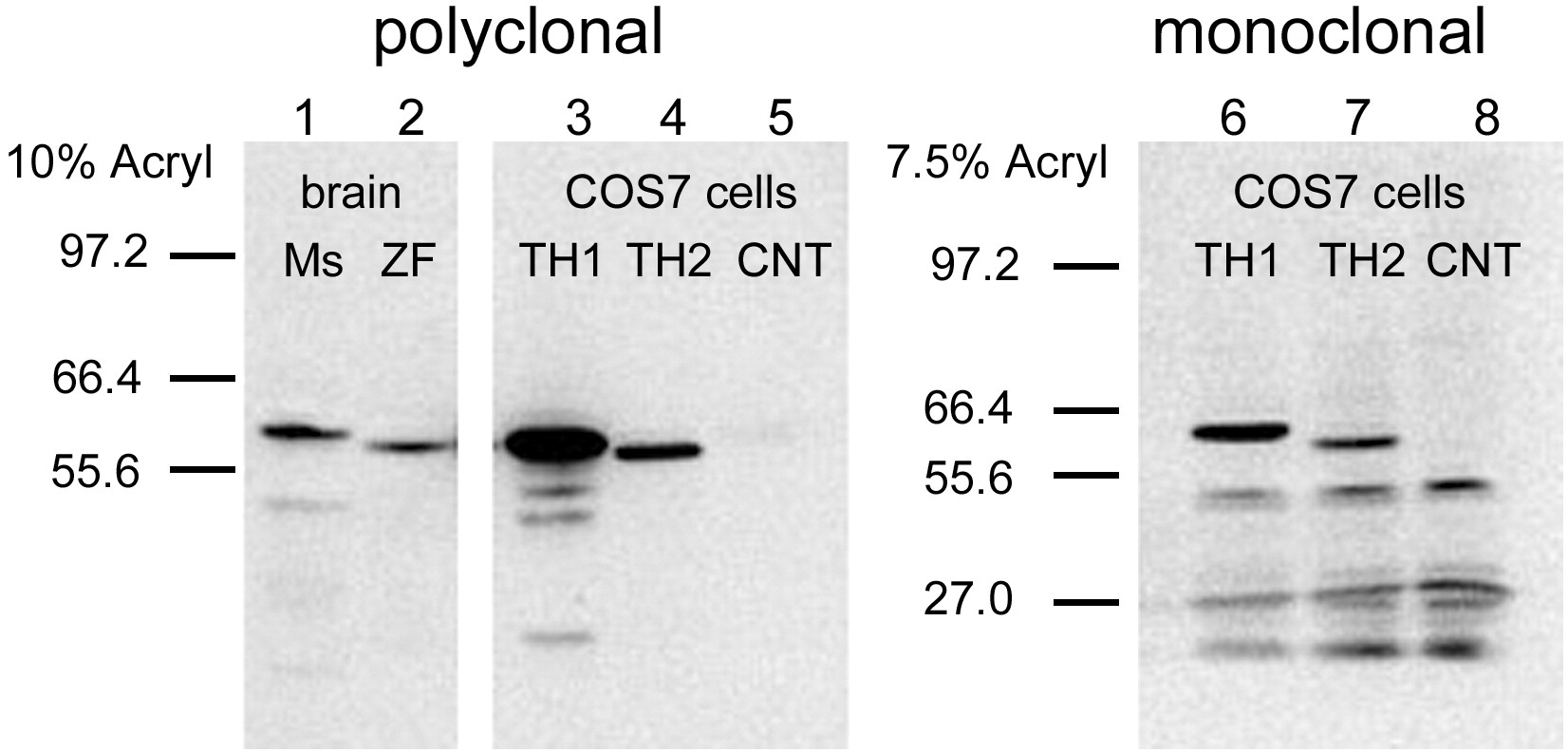
Fig. 3 The specificity of polyclonal (left) and monoclonal (right) anti-TH antibodies for TH1 and TH2 proteins was tested on Western blots. Lanes 1 and 2 correspond to proteins extracted from mouse (Ms) and zebrafish (ZF) brains. Lanes 3 and 6 correspond to zebrafish TH1, and lanes 4 and 7 correspond to TH2, both expressed in COS7 cells. The band of the TH2 shows a lower molecular weight than TH1, and it is labeled much weaker than TH1 by the two antibodies. Lanes 5 and 8 correspond to negative controls (CNT), with lane 5 from untransfected COS7 cells, and lane 8 from COS7 cells transfected with a plasmid in which TH2 is inserted in a reverse orientation. The blot has been overexposed, in order to see the TH2 band better.
Reprinted from Molecular and cellular neurosciences, 43(4), Yamamoto, K., Ruuskanen, J.O., Wullimann, M.F., and Vernier, P., Two tyrosine hydroxylase genes in vertebrates: New dopaminergic territories revealed in the zebrafish brain, 394-402, Copyright (2010) with permission from Elsevier. Full text @ Mol. Cell Neurosci.