
|
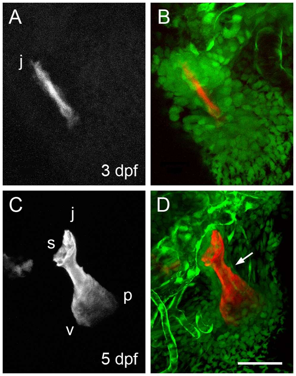
Fig. 6 Arrangements of neural crest-derived mesenchymal cells associated with the opercle developing in the young larva.
Two-color confocal imaging of live preparations. (A, C) Red channel at 3 and 5 dpf showing the Alizarin Red S labeled bone. (B, D) Merge of the red channel and the green channel showing cells expressing the fli1:eGFP transgene. Endothelial cells of capillary tubules also brightly express this transgene. The dense condensation of Op-associated cells present at 3 dpf thins out considerably by 5 dpf, particularly along the very slowing growing jp edge of the bone (arrow in D). Abbreviations and orientations as in Figure 1. Scale bar: 50 μm.