
Fig. 2
- ID
- ZDB-IMAGE-100319-7
- Publication
- Gonzalez-Quevedo et al., 2010 - Neuronal Regulation of the Spatial Patterning of Neurogenesis
- All Figures
- Figures for Gonzalez-Quevedo et al., 2010
|
Fig. 2
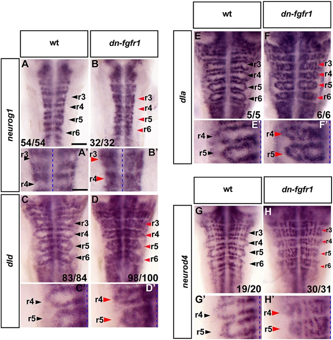
Blocking FGFR Activation Results in Proneural Gene Expression and Differentiating Neurons in Segment Centers
In situ hybridization of 40 hpf zebrafish to detect proneural gene expression (neurog1, dld, dla; [A–F] and [A′–F′]) or differentiating neurons (neurod4; [G and H and G′ and H′]) in either wild-type (wt) or dominant-negative fgfr1 embryos (Tg(hsp70l:dnfgfr1-EGFP)). Heat shock was started at the 22 somite stage. Black arrowheads indicate segment centers; red arrowheads indicate ectopic neurogenesis. (A–H) Scale bar, 50 μm. (A′–H′) Higher-power view of images in (A)–(H); scale bar, 25 μm. Dashed line, midline. See also Figure S1.
Reprinted from Developmental Cell, 18(1), Gonzalez-Quevedo, R., Lee, Y., Poss, K.D., and Wilkinson, D.G., Neuronal Regulation of the Spatial Patterning of Neurogenesis, 136-147, Copyright (2010) with permission from Elsevier. Full text @ Dev. Cell