
Fig. 8
- ID
- ZDB-IMAGE-100319-27
- Publication
- Feng et al., 2010 - Pivotal role of hmx2 and hmx3 in zebrafish inner ear and lateral line development
- All Figures
- Figures for Feng et al., 2010
|
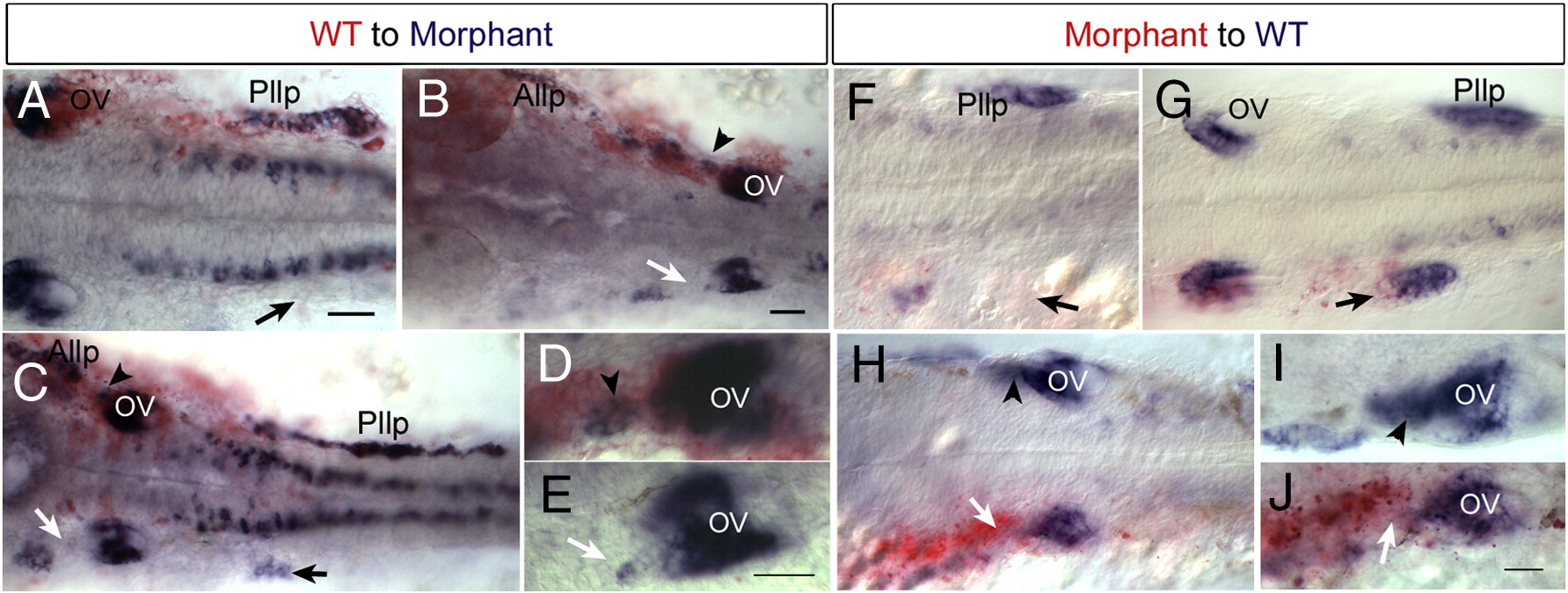
Fig. 8 Cell autonomous effect of Hmx2 and Hmx3 function. Anterior to the left and dorsal view. Donor cells are labeled in red and hmx3 expression in blue. (A–E) Examples of wild-type cells transplanted into hmx2/hmx3 morphant embryos. Arrowheads indicate wild-type cells in morphant backgrounds. Arrows indicated affected tissue in morphant embryos. (F–J) Morphant cells transplanted into wild-type embryos. Arrows indicate morphant donor cells in defective lateral line primordium or otic vesicle. Arrowheads indicate normal development in the control side. Scale bar: 50 μm. Abbreviations: OV, otic vesicle; Pllg, posterior lateral line ganglion; Pllp, posterior lateral primordium; WT, wild type.
Reprinted from Developmental Biology, 339(2), Feng, Y., and Xu, Q., Pivotal role of hmx2 and hmx3 in zebrafish inner ear and lateral line development, 507-518, Copyright (2010) with permission from Elsevier. Full text @ Dev. Biol.