
Fig. 2
- ID
- ZDB-IMAGE-100319-21
- Publication
- Feng et al., 2010 - Pivotal role of hmx2 and hmx3 in zebrafish inner ear and lateral line development
- All Figures
- Figures for Feng et al., 2010
|
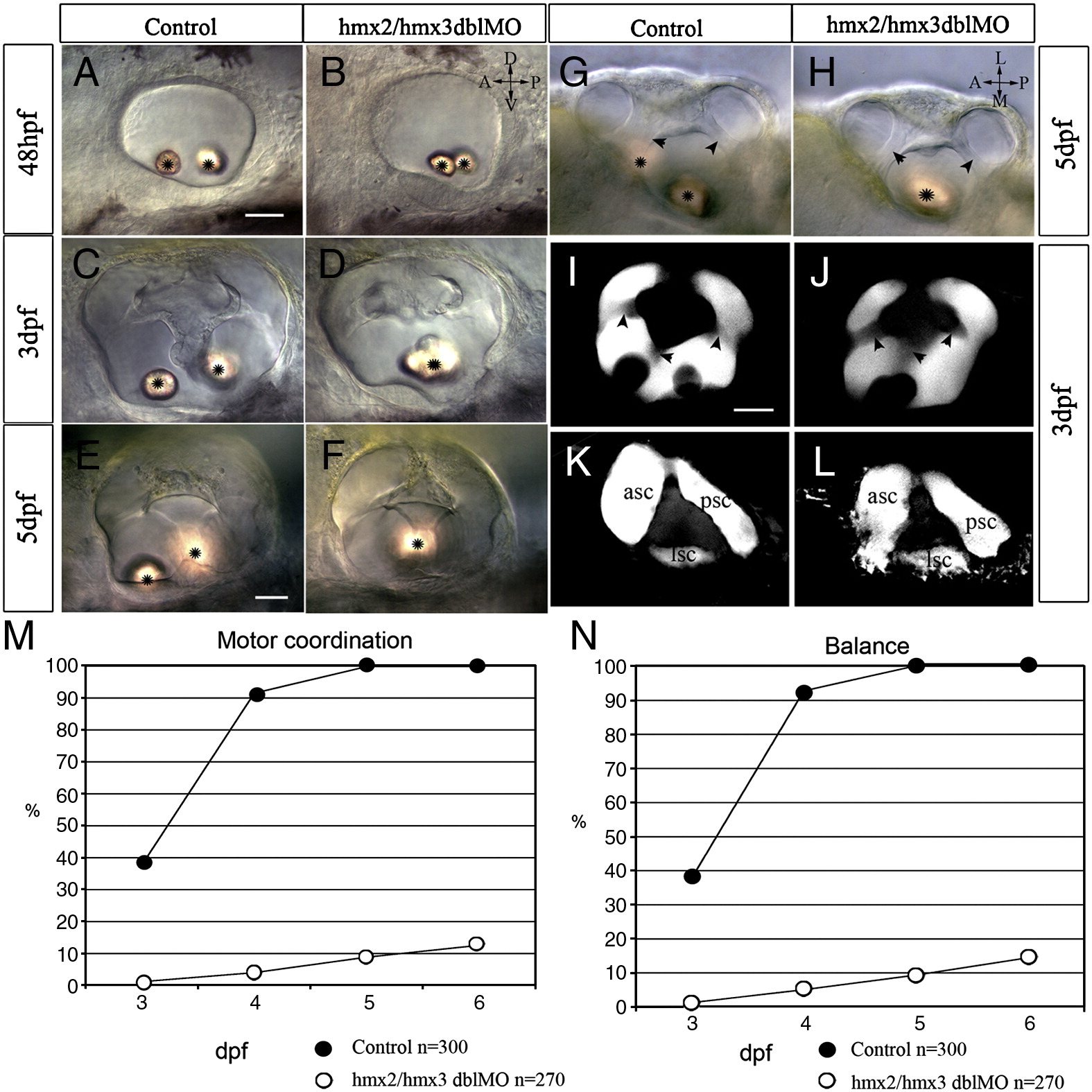
Fig. 2 Ear morphology and vestibular function defects in hmx2/hmx3 morphant embryos. All images are anterior to the left. Bright field DIC images showing normal overall morphology of the ear with fused otoliths (asterisk) in hmx2/hmx3 morphants at the stages indicated (A–H). Lateral view in panels A–F, I–L; dorsal view in panels G, H. Normal semicircular canals in control embryos (I, K) and morphants (J, L) revealed by confocal Z-projection of dye filled lumen. Superficial focal plane in panels I, J, and deeper plane in panels K, L. Arrowheads indicate the lumen of the semicircular canal. Scale bar: 25 μm. Vestibular function is expressed as motor coordination (M) and balance (N) tested for the control embryos and morphants. The x-axis is time (dpf) and the y-axis is percentage of embryos scored positive for the test. Sample size (n = ) is indicated. Abbreviations: ASC, anterior semicircular canal; LSC, lateral semicircular canal; PSC, posterior semicircular canal.
Reprinted from Developmental Biology, 339(2), Feng, Y., and Xu, Q., Pivotal role of hmx2 and hmx3 in zebrafish inner ear and lateral line development, 507-518, Copyright (2010) with permission from Elsevier. Full text @ Dev. Biol.