
Fig. 1
- ID
- ZDB-IMAGE-100211-1
- Publication
- Tay et al., 2010 - The iguana/DZIP1 protein is a novel component of the ciliogenic pathway essential for axonemal biogenesis
- All Figures
- Figures for Tay et al., 2010
|
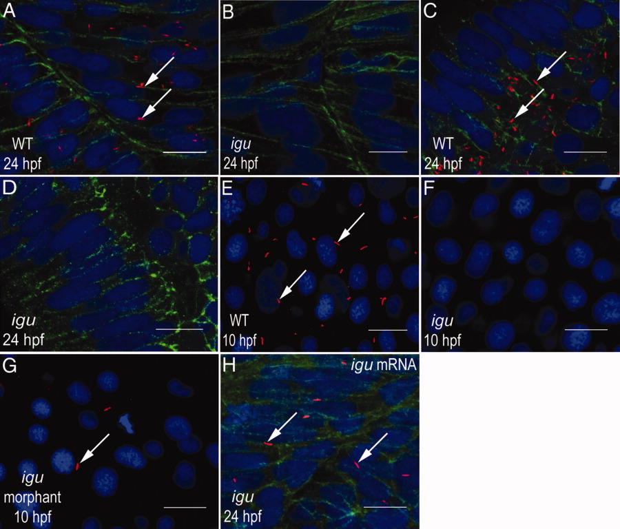
Fig. 1 Igu is essential for primary cilia formation. A: Wild-type myotome showing abundant primary cilia (arrows) on the surface of muscle cells. B: Myotome of an igu mutant embryo showing complete absence of primary cilia. C: Primary cilia (arrows) in differentiating retinal cells of a wild-type embryo. D: Lack of primary cilia from the retina of an igu mutant. E: Primary cilia (arrows) in paraxial mesodermal cells of a wild-type embryo. F: Absence of primary cilia from paraxial mesodermal cells of an igu mutant embryo. G: Severe reduction in numbers of primary cilia (arrow) from paraxial mesodermal cells of an igu morphant embryo. H: An igu mutant embryo injected with igu mRNA showing rescue of the primary cilia (arrows) in muscle cells. Axonemes of primary cilia were visualized with anti-acetylated tubulin antibodies (red), anti-βcatenin antibodies were used to highlight cell membranes (green; A-D and H), and nuclei were visualized with DAPI (blue; 4′,6-diamidine-2-phenylidole-dihydrochloride). In A-D and H, embryos are depicted with anterior to the left and dorsal to the top; E-G show dorsal views of the paraxial mesodermal cells. Scale bars = 10 μm.