
Image
|
Figure Caption
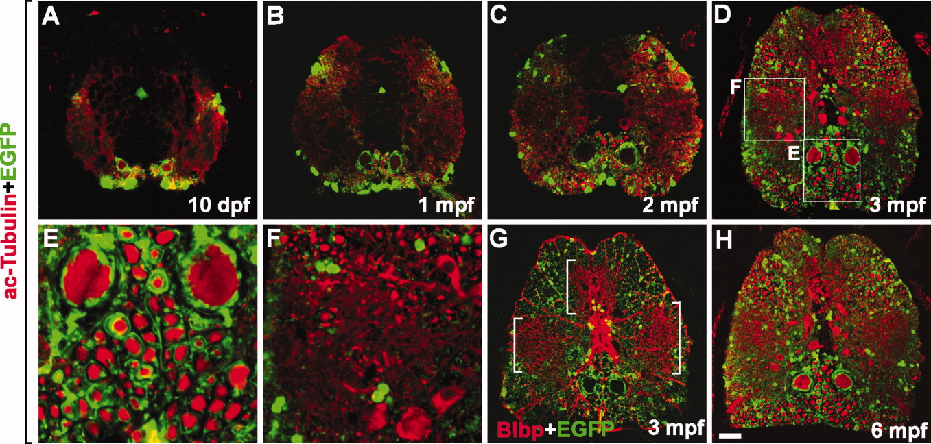
Fig. 4 Axon myelination occurs continuously in the spinal cord of postembryonic zebrafish. All images are transverse sections of the spinal cord of Tg(mbp:egfp) zebrafish, dorsal side up. Stages are indicated on each panel. A-F,H: Labeling with anti-acetylated tubulin antibody to mark axons. E,F: High magnification images of boxed areas in D. G: Labeling with anti-Blbp antibody to mark radial glia. Bracketed areas indicate clusters of highly branched radial glial processes. Scale bars = 25 μm in A, 50 μm in B, 80 μm in C, 100 μm in D,G,H, 25 μm in E,F.
Acknowledgments
This image is the copyrighted work of the attributed author or publisher, and
ZFIN has permission only to display this image to its users.
Additional permissions should be obtained from the applicable author or publisher of the image.
Full text @ Dev. Dyn.