
IMAGE
Fig. 6
- ID
- ZDB-IMAGE-100209-10
- Publication
- Siddiqui et al., 2010 - The tight junction component Claudin E is required for zebrafish epiboly
- All Figures
- Figures for Siddiqui et al., 2010
Image
|
Figure Caption
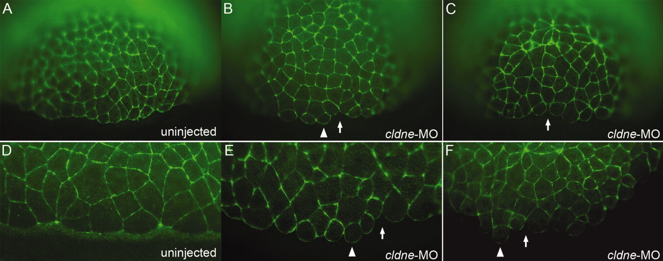
Fig. 6 Phalloidin staining reveals abnormal marginal enveloping layer (EVL) cell morphology. A-F: Lateral views of phalloidin stained embryos, injected constructs indicated in lower right. Arrowheads indicate abnormally round marginal EVL cells; arrows indicate gaps between cells. A-C: Embryos at dome to 30% epiboly stage. D-F: Embryos at shield stage.
Figure Data
Acknowledgments
This image is the copyrighted work of the attributed author or publisher, and
ZFIN has permission only to display this image to its users.
Additional permissions should be obtained from the applicable author or publisher of the image.
Full text @ Dev. Dyn.