
Fig. 7
- ID
- ZDB-IMAGE-100128-22
- Publication
- Mitchell et al., 2010 - Effect of Vascular Cadherin Knockdown on Zebrafish Vasculature during Development
- All Figures
- Figures for Mitchell et al., 2010
|
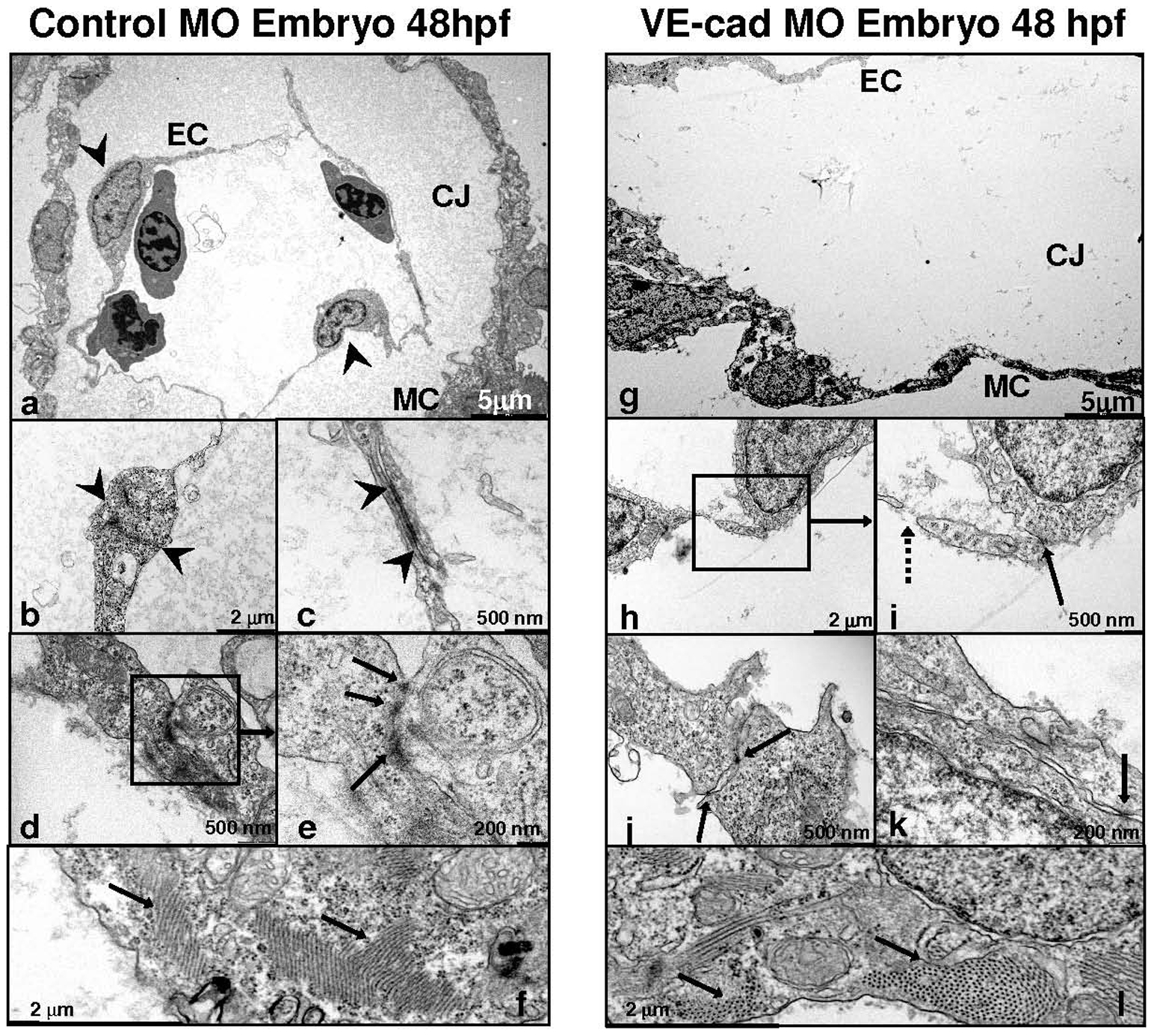
Fig. 7 Loss of VE-cadherin produces defective endocardial junctions and increased permeability:
Images obtained by transmission electron microscopy of control (a–f) and VE-cad knockdown (g–l) embryos at 48 hpf. The atria of control embryos (a) demonstrate a modest layer of cardic jelly (CJ) between the endocardial (EC) and myocardial layers (MC) (arrowheads are endothelial cell nuclei in a). Knockdown embryos (g) show a stretched myocardium, wide endocardial/myocardial separation and decreased electron density of the cardiac jelly. Mature-appearing, long endocardial junctions are present in controls (between arrowheads, b and c; arrows e). VE-cad MO embryos have smaller, fewer, less well-developed junctions (solid arrows i–k) and endothelial gaps (dashed arrow, i). Both control and knockdown embryos (f and l) demonstrate similar overall numbers of well-developed contractile elements in their myocardial layers (f and l).