
|
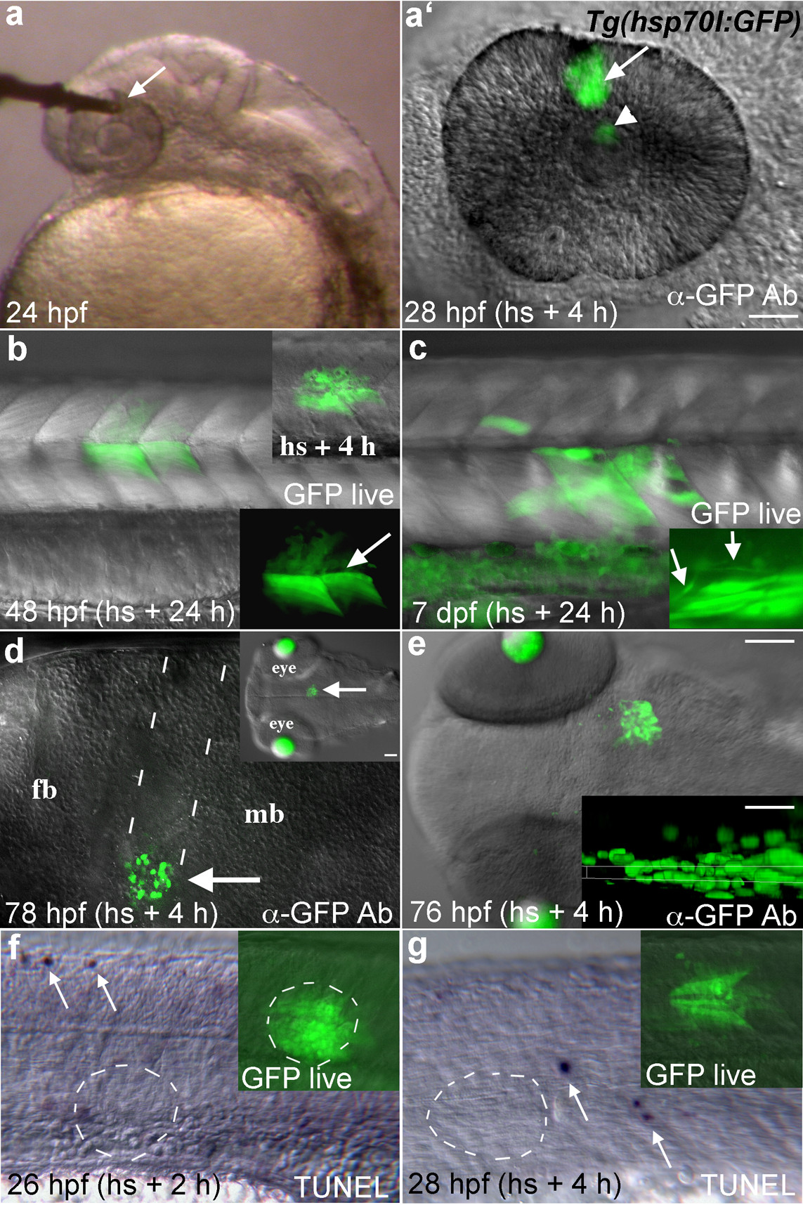
Fig. 2 GFP expression in Tg(hsp70l:GFP) embryos following local heat shock. (a) A 50 μm diameter microheater tip (arrow) touching the eye of a 24 hpf embryo mounted in low melting temperature agarose. (a′) GFP expressing cells (arrow) were seen in the heated region 4 hours later. GFP fluorescence is also seen reflected by the lens (arrowhead). (b) GFP expression (arrows) in a 48 hpf transgenic embryo heat shocked 24 h earlier in the trunk with a 100 μm optical fiber tip. Muscle fiber morphology in GFP expressing cells is normal (arrow in lower inset). Upper inset shows GFP expression 4 h after heat shock. (c) Transgene activation in a 7 dpf larva that was heat shocked at 6 dpf. Inset shows normal spinal neuron morphology (arrows) in a different individual. (d) A 50 μm tip was pushed into the brain of this embryo at 72 hpf to locally heat deep tissue (arrows, dotted lines show entry pathway). Lateral view of the brain, dorsal up. Transgene expression was activated with relatively little damage from the optical fiber. Inset shows the region of gene activation in a dorsal view. Lens tissue fluoresces in this transgenic line at these later developmental stages. (e) Dorsal view of GFP expression in the midbrain of a 76 hpf larva 4 hours after heat shock. Inset shows a resliced (xz) view of the GFP expression domain (maximum intensity projection along the y axis), dorsal up. GFP expression was activated in cells ∼30 μm away from the dorsal surface that contacted the fiber optic tip. (f, g) TUNEL labeling in the trunk region 2 and 4 hours after heat shock, arrows indicate labeled apoptotic cells. No increased cell death was seen in the area of local heat shock (dashed circles). Insets show GFP expression just prior to fixation. Lateral views. Scale bars: a-d, f, g, 50 μm; e, 100 μm, inset, 30 μm.