
|
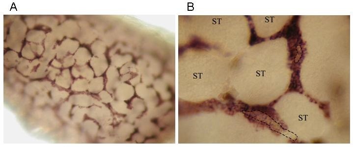
Fig. 7 Whole mount in situ hybridization of insl3 on zebrafish testis. A) Overview of the positive insl3 in situ hybridization signal in zebrafish testis, clearly showing positive insl3 in situ hybridization signal in the interstitial area. B) Detailed view of A, showing that only the cytoplasm of the Leydig cells in zebrafish testis shows the positive in situ hybridization signal. Blood vessels (encircled by dashes) containing erythrocytes are often visible in the Leydig cell clusters. The seminiferous tubules (ST), containing Sertoli cells and germ cells in different stages of spermatogenesis, remain completely unstained.