
Fig. 4
- ID
- ZDB-IMAGE-100114-27
- Antibodies
- Publication
- Codina et al., 2010 - Loss of Smyhc1 or Hsp90alpha1 function results in different effects on myofibril organization in skeletal muscles of zebrafish embryos
- All Figures
- Figures for Codina et al., 2010
|
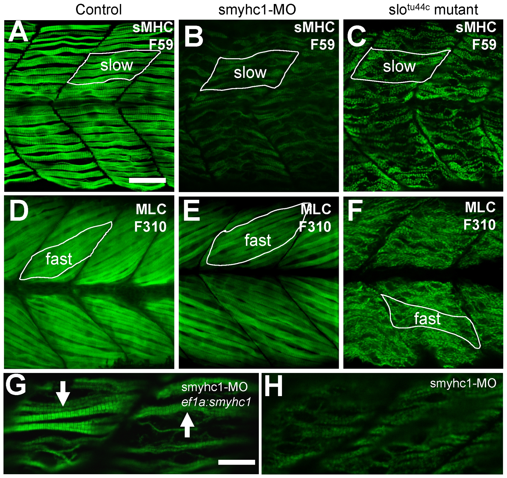
Fig. 4 Effects of smyhc1 knockdown or hsp90α1 mutation on myosin thick filament organization in skeletal muscles of zebrafish embryos.
A–C. Anti-MyHC antibody (F59) staining shows the organization of thick filaments in trunk slow muscles of control (A), smyhc1 knockdown (B), or slotu44c mutant (C) embryos at 48 hpf. D–F. Anti-MLC antibody (F310) staining shows the organization of thick filaments in trunk fast muscles of control (D), smyhc1 knockdown (E), or slotu44c mutant (F) embryos at 72 hpf. Note, fast fibers project with a 30 degree angle with respect to the axial structure, whereas slow fibers project in parallel to the axial structure. G, H. Anti-MyHC antibody (F59) staining shows the rescue of thick filaments in smyhc1 knockdown zebrafish embryos co-injected with ef1a:smyhc1 DNA construct (G), or ATG-MO alone (H). Scale bar = 25 μm in A, 10 μm in G.