
Image
|
Figure Caption
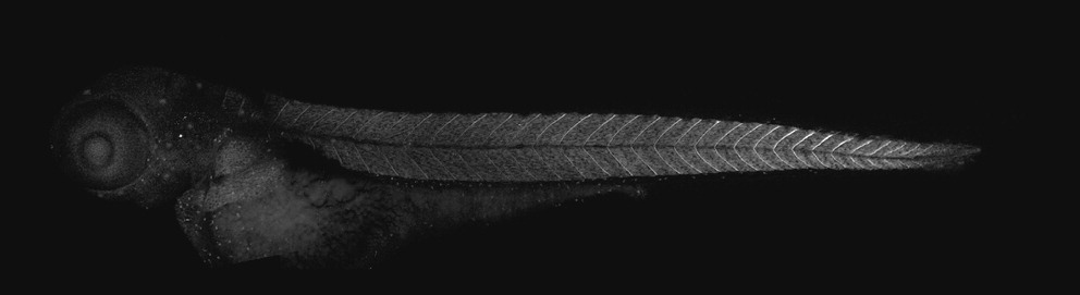
Fig. 4 Immunostaining of MMP11 protein in a 48-hpf zebrafish embryo. Projections of confocal stacks were assembled to show the complete staining pattern of anti-zebrafish MMP11 in a 48-hpf embryo. Strong labeling is detected at maturing somite boundaries. Individual migrating mesenchymal cells are also labeled throughout the head and trunk (anterior to the left).
Acknowledgments
This image is the copyrighted work of the attributed author or publisher, and
ZFIN has permission only to display this image to its users.
Additional permissions should be obtained from the applicable author or publisher of the image.
Full text @ Zebrafish