
Image
|
Figure Caption
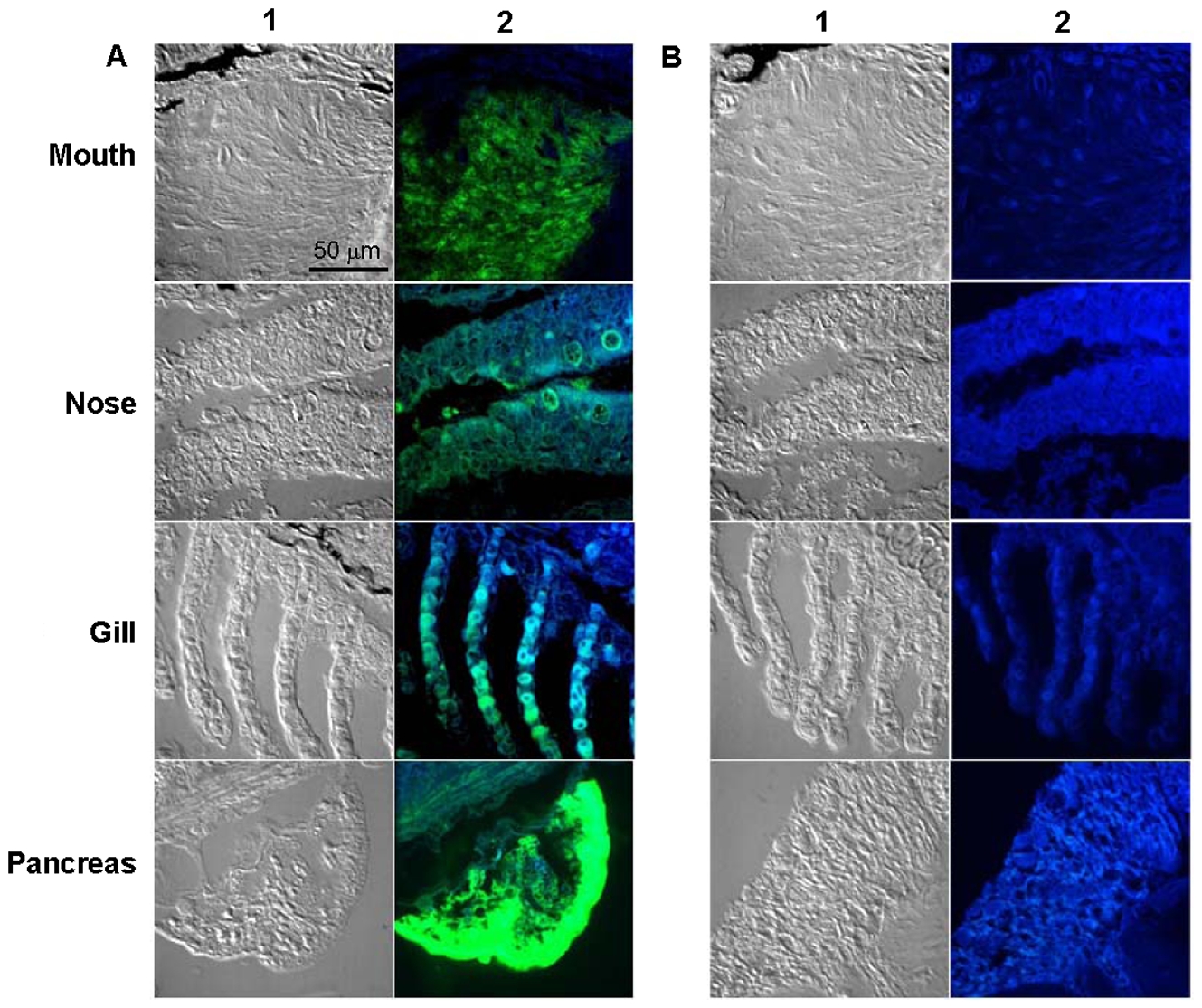
Fig. 3 Detection of trypsin by confocal microscopy.
Confocal DIC images (a, 60X and b, 100X) of zebrafish sections of mouth, nose, gill and pancreas stained by immunohistochemistry using primary antibody (a) PRAHT, (b) Non-immunized rabbit IgG. Alexa Fluor 488 goat anti-rabbit IgG was used as secondary antibody. We used two channel image acquisition to examine the expression of trypsins that were probed with Alexa Fluor. A 488 nm and a 405 nm laser were used to excite the Alexa Fluor (Green) and autofluorescence (Blue), respectively. Columns 1 and 2 show brightfield and fluorescence images respectively.
Figure Data
Acknowledgments
This image is the copyrighted work of the attributed author or publisher, and
ZFIN has permission only to display this image to its users.
Additional permissions should be obtained from the applicable author or publisher of the image.
Full text @ PLoS One