
Fig. S6
- ID
- ZDB-IMAGE-100107-11
- Publication
- Soyer et al., 2010 - Rfx6 is an Ngn3-dependent winged helix transcription factor required for pancreatic islet cell development
- All Figures
- Figures for Soyer et al., 2010
|
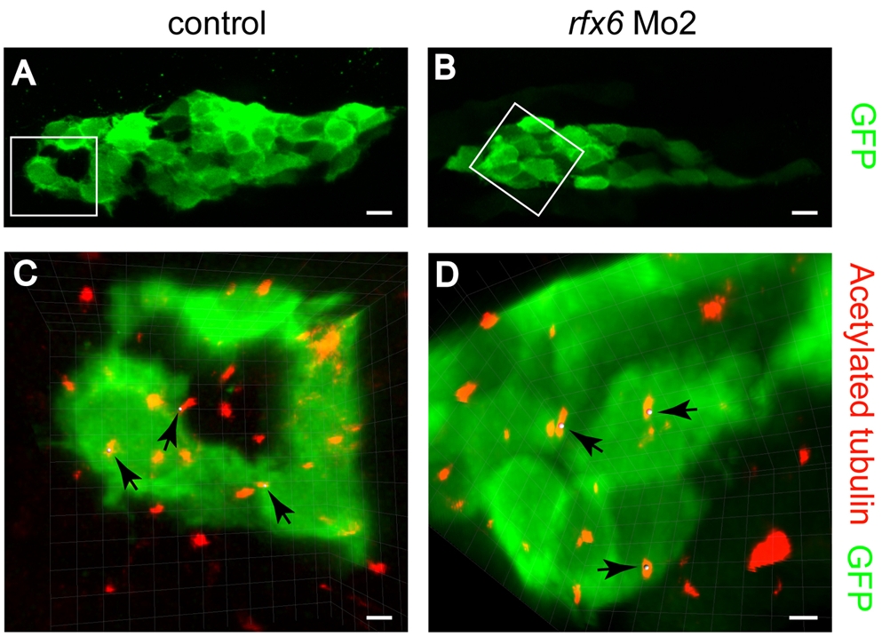
Fig. S6 Acetylated-tubulin labelling reveals ciliated cells within the developing pancreas of wild-type and morphant zebrafish. Experiments to visualize primary cilia were performed on transgenic zebrafish embryos tg(pax6:GFP) where pancreatic endocrine cells can be detected by GFP expression (Delporte et al., 2008). (A,B) Pancreatic cell labelling using the tg(pax6:GFP) transgenic line underlines the developing pancreas in control (A) and rfx6 Mo2 morphants (B). (C,D) Magnifications of the area in white boxes in A and B with the acetylated-tubulin-labelled cilia in red and pax6:GFP in green. Arrows in C and D point to cilia. The same cilia are marked with a grey dot visible in Movies 1 and 2. Scale bars: 10 mm in A,B; 2 mm in C,D.