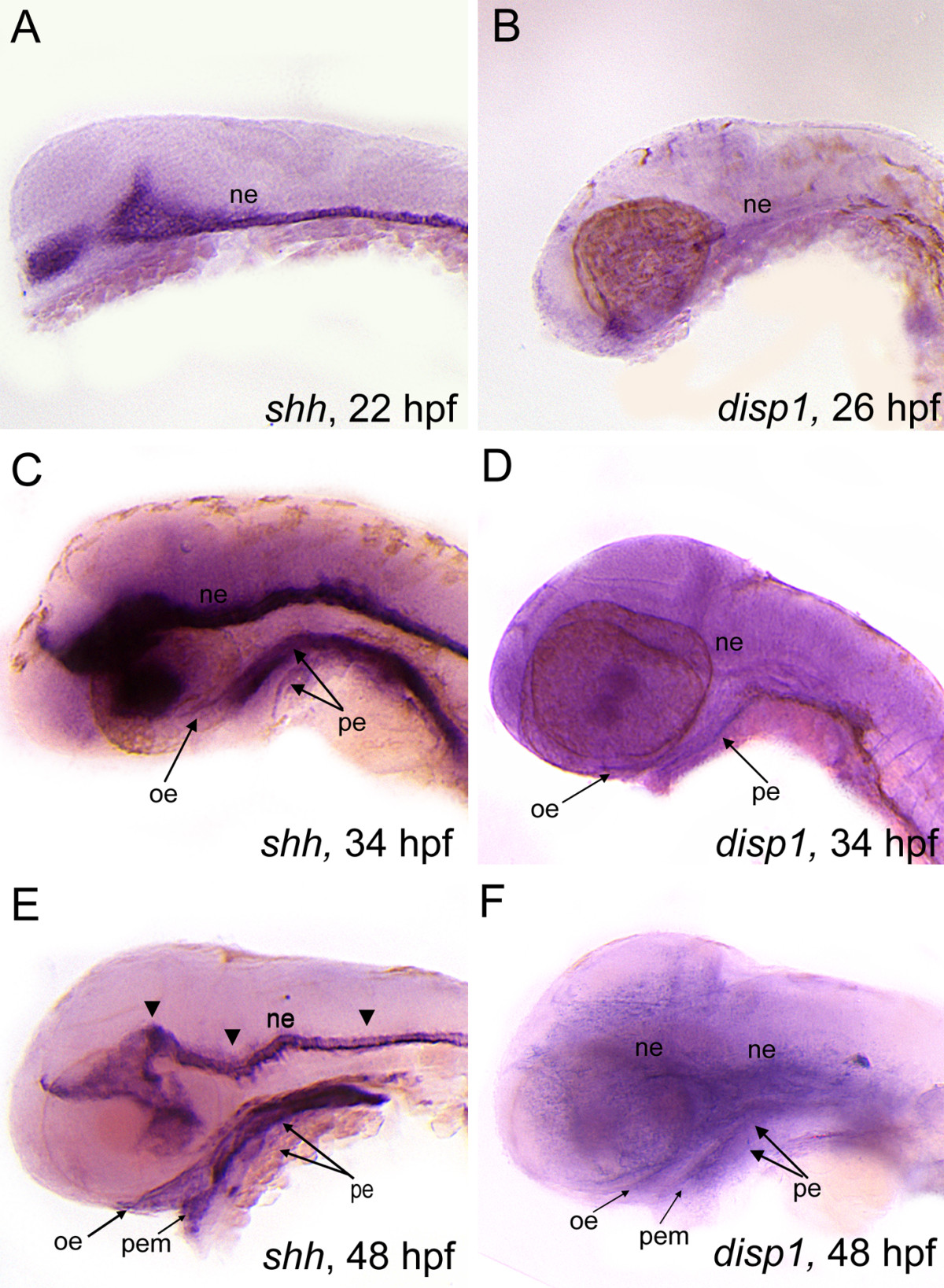
Fig. 7 shh and disp1 are coexpressedin the developing head. Lateral views of wild type embryos labeled with RNA probe for shh (A,C,E) or disp1 (B,D,F). (A,B) At 22 hpf (shh) - 25 hpf (disp1)are both expressed in ventral neuroectoderm (ne). (C,D) At 34 hpf, shh and disp1 expression becomes detectable in oral ectoderm (oe) and pharyngeal endoderm (pe), in addition to the neuroectoderm (ne). Arrowheads denote expression within the neuroectoderm for both shh and disp1. (E-F) By 48 hpf, shh and disp1 expression becomes more prominent in the oe and pe, and is further expanded to the pharyngeal ectodermal margin (pem). shh and disp1 expression persists in the ne at 48 hpf.
Image
Figure Caption
Figure Data
Acknowledgments
This image is the copyrighted work of the attributed author or publisher, and
ZFIN has permission only to display this image to its users.
Additional permissions should be obtained from the applicable author or publisher of the image.
Full text @ BMC Dev. Biol.

