
|
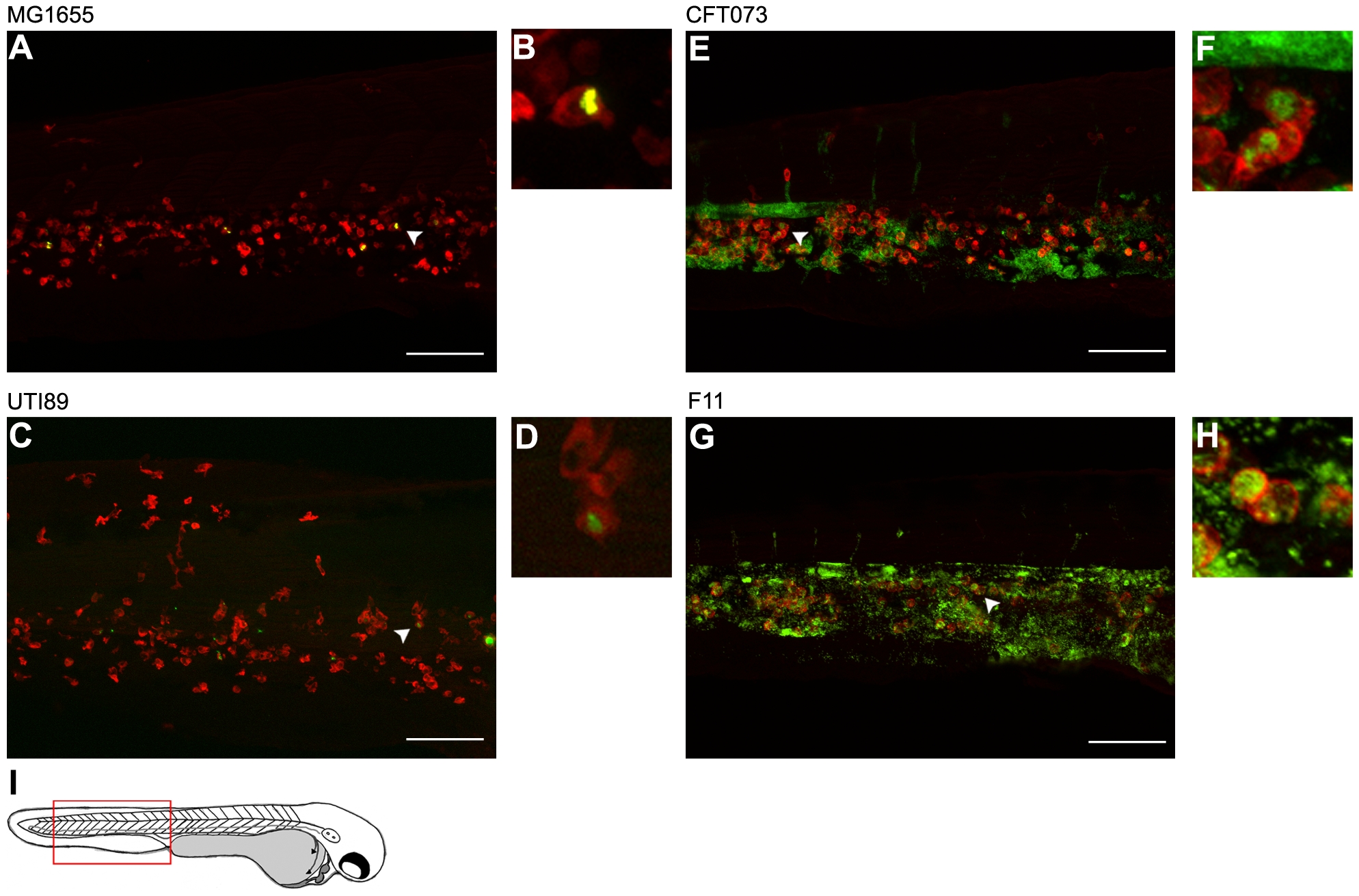
Fig. 7 Differential growth and phagocytosis of E. coli isolates within the blood.
Zebrafish embryos were infected via the blood with 4,000–6,500 CFU of (A and B) MG1655, (C and D) UTI89, (E and F) CFT073, or (G and H) F11. All bacterial strains carry pGEN-GFP(LVA) for constitutive expression of destabilized GFP (green). At 12 hpi, samples were fixed and phagocytes (red) were labeled using L-plastin-specific antibody for visualization by fluorescent confocal microscopy. Regions highlighted by arrowheads in (A), (C), (E), and (G) are shown further magnified in panels (B), (D), (F), and (H), respectively. All images shown are representative of the pool of embryos imaged. MG1655- and UTI89-infected embryos were viable and healthy in appearance prior to sacrifice for microscopy, whereas fish inoculated with CFT073 or F11 were notably sick and near death at time of collection. Scale bars = 100 μm. (I) Diagram of a 48 hpf embryo with the region imaged in (A), (C), (E), and (G) denoted by a red box.