
|
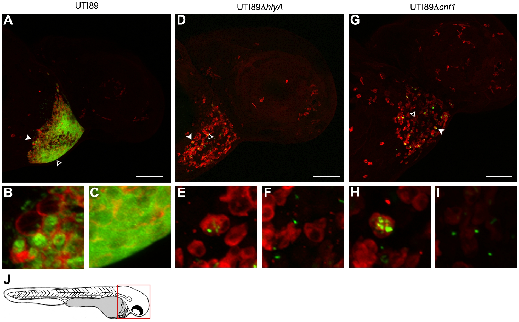
Fig. 5 Phagocyte localization and bacterial internalization within the pericardial cavity.
Zebrafish embryos were injected via the P.C. with 4,000–6,500 CFU of (A–C) wt UTI89, (D–F) UTI89ΔhlyA, or (G–I) UTI89Δcnf1, each carrying pGEN-GFP(LVA) for constitutive expression of destabilized GFP protein (green). At 6 hpi, samples were fixed and processed for fluorescent confocal microscopy, using anti-L-plastin antibody to label phagocytes (red). Examples of internalized (solid arrowheads) and free extracellular (hollow arrowheads) bacteria in (A, D, and G) are shown at higher magnification in (B, E, and H) and (C, F, and I), respectively. Representative images are shown. All fish were viable at the time of sacrifice. Scale bars = 100 μm. (J) Diagram of a 48 hpf embryo with the area imaged in (A), (D), and (G) highlighted by a red box.