
|
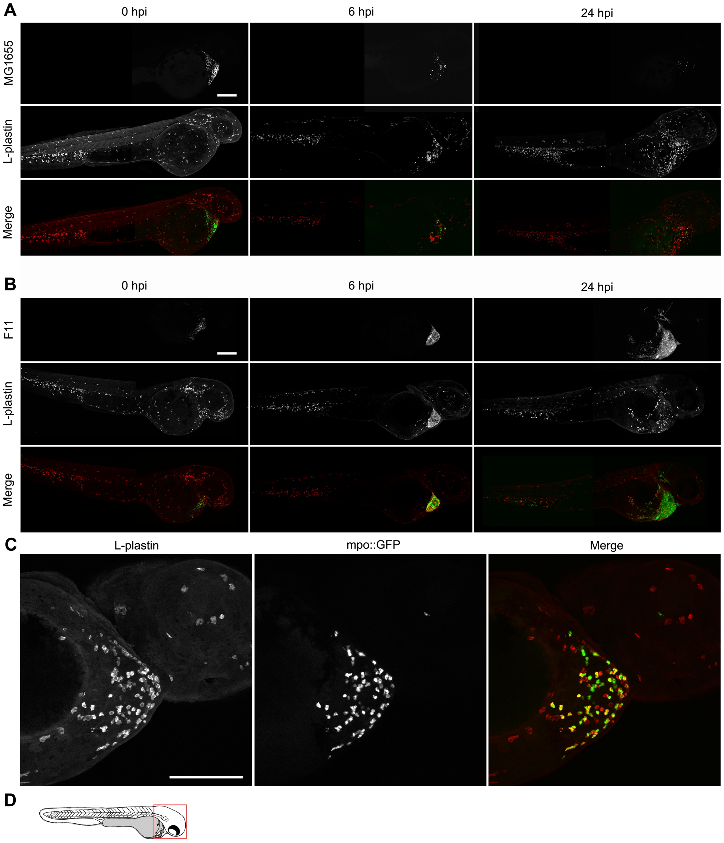
Fig. 4 Phagocyte recruitment and abatement within the zebrafish pericardial cavity.
(A and B) Zebrafish embryos were inoculated via the P.C. with 4,000–6,500 CFU of (A) the K12 strain MG1655 or (B) the ExPEC isolate F11, both carrying pGEN-GFP(LVA) for constitutive expression of destabilized GFP protein (green). Samples were fixed at 0, 6, and 24 hpi and processed for fluorescent confocal microscopy. Phagocytes (red) were detected using L-plastin-specific antibody. For clarity, the merged image at each time point is accompanied by images showing only corresponding single channel signals from bacteria or L-plastin. Each embryo was visualized by stitching together two z-projections generated from 40 to 50 5-μm-thick optical sections. Scale bar = 200 μm. (C) Tg(mpo::GFP) zebrafish embryos, in which GFP expression is under control of the neutrophil-specific MPO promoter, were injected via the P.C. with ∼5,000 CFU of the ExPEC isolate F11. At 4 hpi, samples were fixed and stained using anti-L-plastin antibody to label total phagocytes (red) relative to the GFP-positive neutrophils (green plus red). Bacteria present within the P.C. are not shown. Scale bar = 100 μm. (D) Diagram of a 48 hpf embryo with the area imaged in (C) highlighted by a red box. Embryos shown here are representative. All fish were viable at the time of sacrifice, except the 24 hpi F11-infected fish, which died prior to collection.