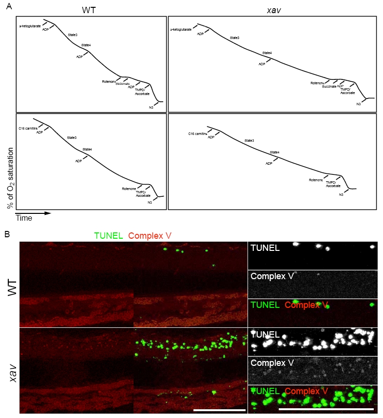
Fig. S4 xav mutants exhibit respiratory deficiency. A. Polarographic traces showing O2 consumption by mitochondria in WT and xav homogenates. Freshly prepared homogenates from WT and xav embryos were incubated in an oxygen sensor chamber, and O2 consumption (y axis) as a function of incubation time (x axis) was recorded. In the upper panels, homogenates were incubated with α-ketoglutarate + malate, and in the lower panels, homogenates were incubated with fatty acid (C16 carnitine + malate). Maximal rates of electron transfer were determined from the rates of O2 consumption driven by ADP (0.2 mM) and inorganic phosphate (state 3) and state 4 determined from the rate of O2 consumption upon conversion of the ADP to ATP. N = >10 embryos from at least 2 carrier pairs for each metabolic assay. B. At ∼56 hpf, a higher level of F1-F0 ATPase (complex V) protein was detected by immunostaining (red) in xav mutants (lower panels) compared to WT embryos (upper panels). A higher magnification view of the spinal cord is shown in the right most panels. TUNEL staining (green) was performed simultaneously and showed that there is increased cell death in the spinal cord in xav and that complex V positive cells are also TUNEL positive in xav. N = >10 embryos from at least 2 carrier pairs for each immunostaining assay. Scale bar = 100 μm.
Image
Figure Caption
Figure Data
Acknowledgments
This image is the copyrighted work of the attributed author or publisher, and
ZFIN has permission only to display this image to its users.
Additional permissions should be obtained from the applicable author or publisher of the image.
Full text @ PLoS One

