
IMAGE
Fig. S4
- ID
- ZDB-IMAGE-091217-130
- Publication
- Edeling et al., 2009 - Structural requirements for PACSIN/Syndapin operation during zebrafish embryonic notochord development
- All Figures
- Figures for Edeling et al., 2009
Image
|
Figure Caption
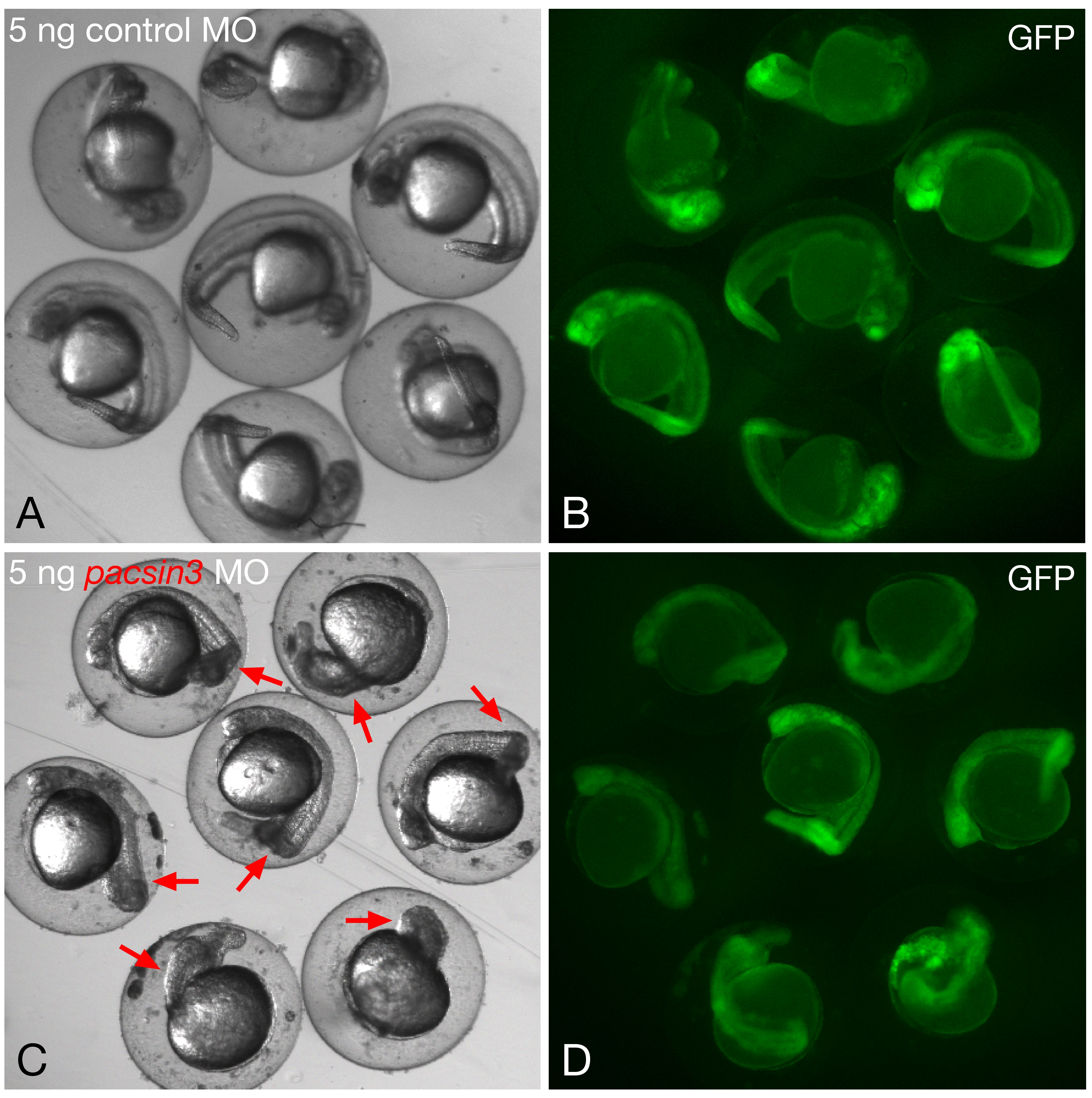
Fig. S4 Phenotypic range with 5 ng pacsin3 MO. (A–B) Gross morphology of embryos within the chorion at 24 hpf after injection of 5 ng control MO and 50 pg GFP cRNA at the one- to two-cell stage. Bar = 250 μm. (C–D) Gross morphology of embryos within the chorion at 24 hpf after injection of 5 ng pacsin3 MO and 50 pg GFP cRNA at the one- to two-cell stage. Red arrows indicate obvious morphological abnormalities in the morphant embryos. Note too the generally reduced anterioposterior axial length in the pacsin3 MO-injected embryos.
Acknowledgments
This image is the copyrighted work of the attributed author or publisher, and
ZFIN has permission only to display this image to its users.
Additional permissions should be obtained from the applicable author or publisher of the image.
Full text @ PLoS One