
Fig. 4
- ID
- ZDB-IMAGE-091215-65
- Publication
- Lee et al., 2009 - Notch Signaling Functions as a Cell-Fate Switch between the Endothelial and Hematopoietic Lineages
- All Figures
- Figures for Lee et al., 2009
|
Fig. 4 Notch Signaling Functions as a Cell-Fate Switch between the Endothelial and the Hematopoietic Lineages in the Nascent Mesoderm
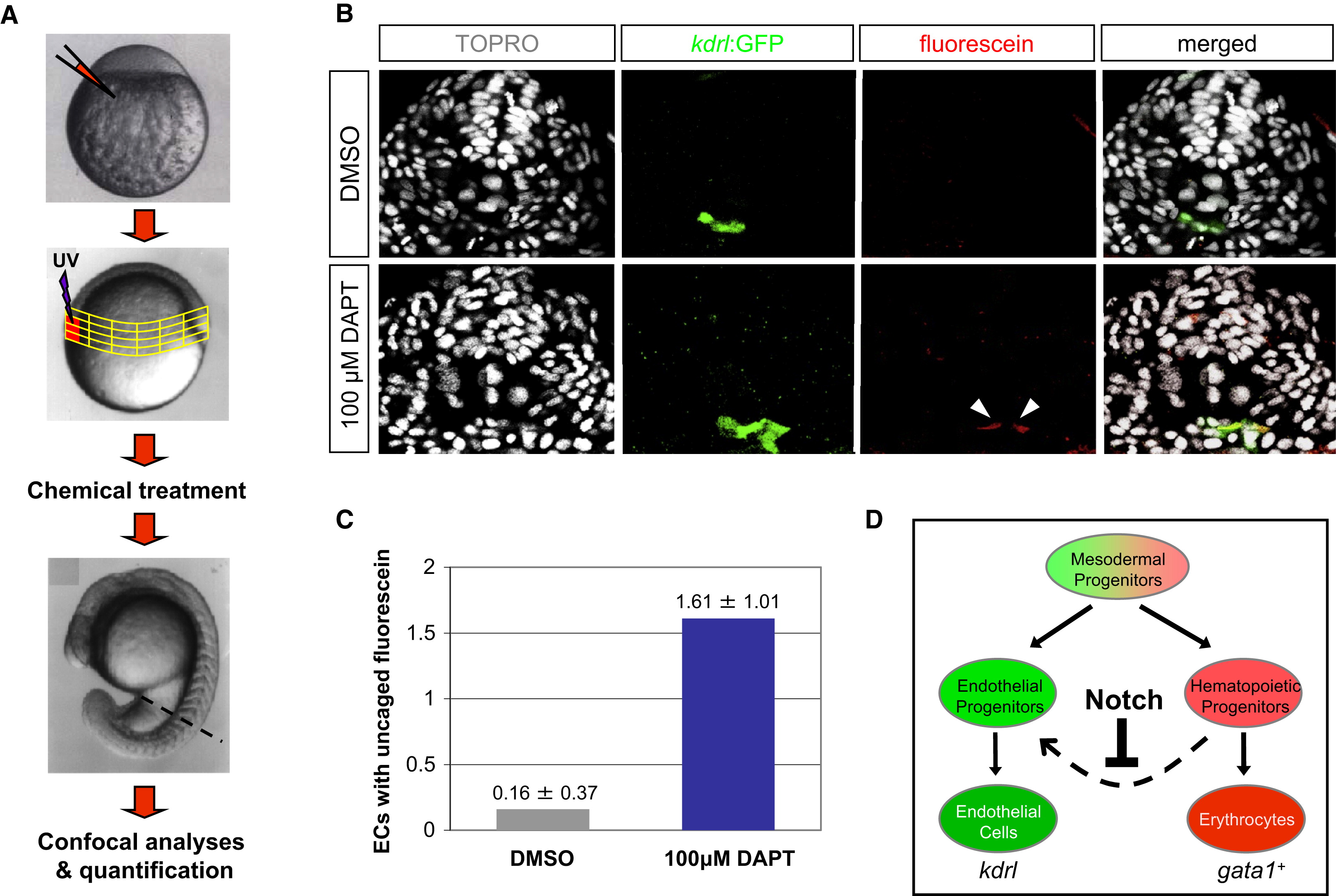
(A) Experimental scheme of the lineage-tracing experiment. Single-cell-stage embryos were injected with DMNB-caged fluorescein-conjugated dextran. At shield stage, ten cells within the ventral margin, which predominantly gives rise to hematopoietic cells, were uncaged with UV irradiation. Subsequently, embryos were treated with either DMSO or DAPT until 18 hpf.
(B) Transverse sections of 18 hpf embryos treated with DMSO or DAPT after uncaging, visualized for TOPRO (white), kdrl:GFP (green), and uncaged fluorescein (red). White arrowheads point to endothelial cells that contain uncaged fluorescein in DAPT-treated embryos.
(C) Quantification of the number of endothelial nuclei with uncaged fluorescein per focal plane in embryos treated with DMSO (n = 18) or DAPT (n = 13). Embryos treated with DAPT contained 1.61 (s = 1.01) fluorescein-positive endothelial cells, in comparison to 0.16 (s = 0.37) in DMSO-treated embryos.
(D) Our proposed model: Notch signaling negatively regulates endothelial cell number by limiting the mesodermal cells that can differentiate into endothelial progenitors. Attenuated Notch activity causes mesodermal cells with hematopoietic potential to differentiate into endothelial progenitors, thus increasing the number of endothelial cells.