
Fig. 3
- ID
- ZDB-IMAGE-091215-64
- Genes
- Publication
- Lee et al., 2009 - Notch Signaling Functions as a Cell-Fate Switch between the Endothelial and Hematopoietic Lineages
- All Figures
- Figures for Lee et al., 2009
|
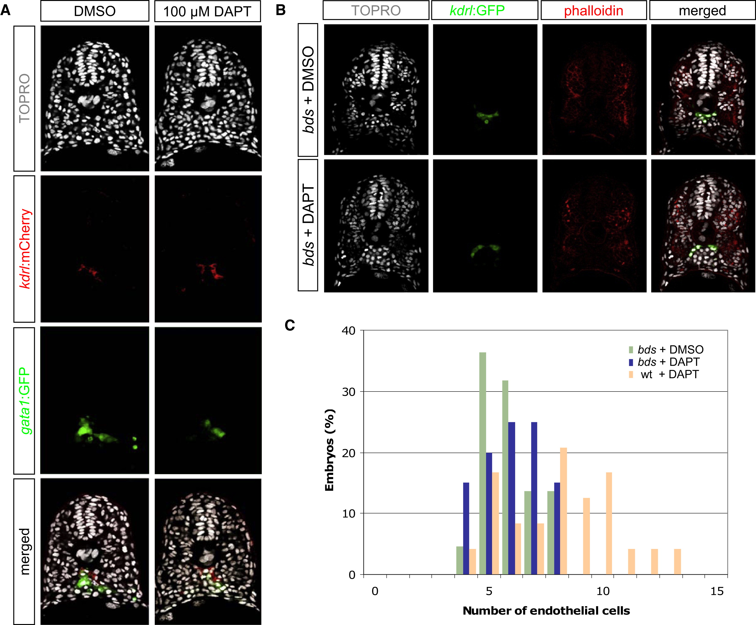
Fig. 3 Reduced Notch Activity Causes an Increase of Endothelial Cells at the Expense of Hematopoietic Cells
(A) Transverse sections of 18 hpf DMSO- or DAPT-treated embryos, visualized for TOPRO (white), kdrl:mCherry (red), and gata1:GFP (green). DAPT-treated embryos exhibited a concomitant gain of endothelial cells and loss of hematopoietic cells.
(B) Transverse sections of 20 hpf DMSO- or DAPT-treated phenotypic bloodless (bds) mutant embryos, visualized for TOPRO (white), kdrl:GFP (green), and phalloidin (red).
(C) Quantification of the number of endothelial nuclei per focal plane in DMSO-treated bds (n = 22), DAPT-treated bds (n = 20), and DAPT-treated wild-type sibling (n = 24) embryos. DAPT treatment in embryos lacking primitive hematopoietic cells (bds + DAPT) did not result in a change in the number of endothelial cells, suggesting that the ectopic endothelial cells observed in DAPT-treated wild-type embryos might have originated from cells that normally produce hematopoietic cells.