
Fig. S7
- ID
- ZDB-IMAGE-091215-60
- Publication
- Lee et al., 2009 - Notch Signaling Functions as a Cell-Fate Switch between the Endothelial and Hematopoietic Lineages
- All Figures
- Figures for Lee et al., 2009
|
Fig. S7
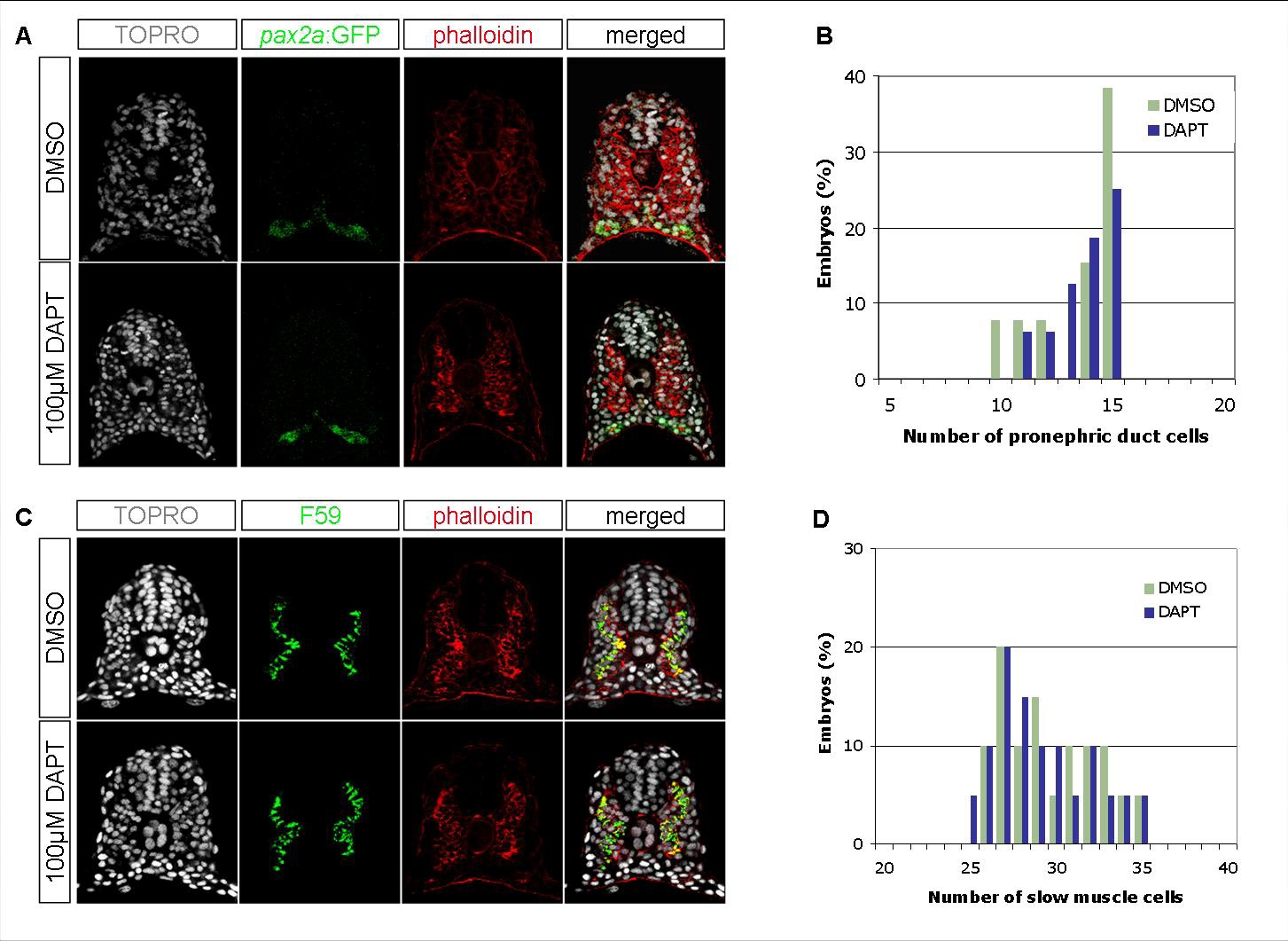
Notch Signaling Does Not Affect the Number of Other Nonaxial Mesoderm-Derived Lineages
(A) Transverse sections of 18 hpf embryos, DMSO- or DAPT-treated, visualized for TOPRO (white), pax2a:GFP (green), and phalloidin (red). (B) Quantification of pronephric duct cell nuclei per focal plane in DMSO-treated (n=13) or DAPT-treated (n=16) embryos. (C) Transverse sections of 18 hpf embryos, DMSO- or DAPT-treated, visualized for TOPRO (white), S59 antibody (green), and phalloidin (red). (D) Quantification of slow muscle fiber cell nuclei per focal plane in DMSO-treated (n=20) or DAPT-treated (n=20) embryos. Embryos treated with DAPT contained 12.50 (s=1.63) pronephric duct cell nuclei and 29.20 (s=2.84) slow muscle fiber cell nuclei per section, which were comparable to DMSO-treated embryos (12.23 (s=2.05) pronephric duct cell nuclei and 29.70 (s=2.77) slow muscle fiber cell nuclei).