
IMAGE
Fig. S6
- ID
- ZDB-IMAGE-091215-59
- Publication
- Lee et al., 2009 - Notch Signaling Functions as a Cell-Fate Switch between the Endothelial and Hematopoietic Lineages
- All Figures
- Figures for Lee et al., 2009
Image
|
Figure Caption
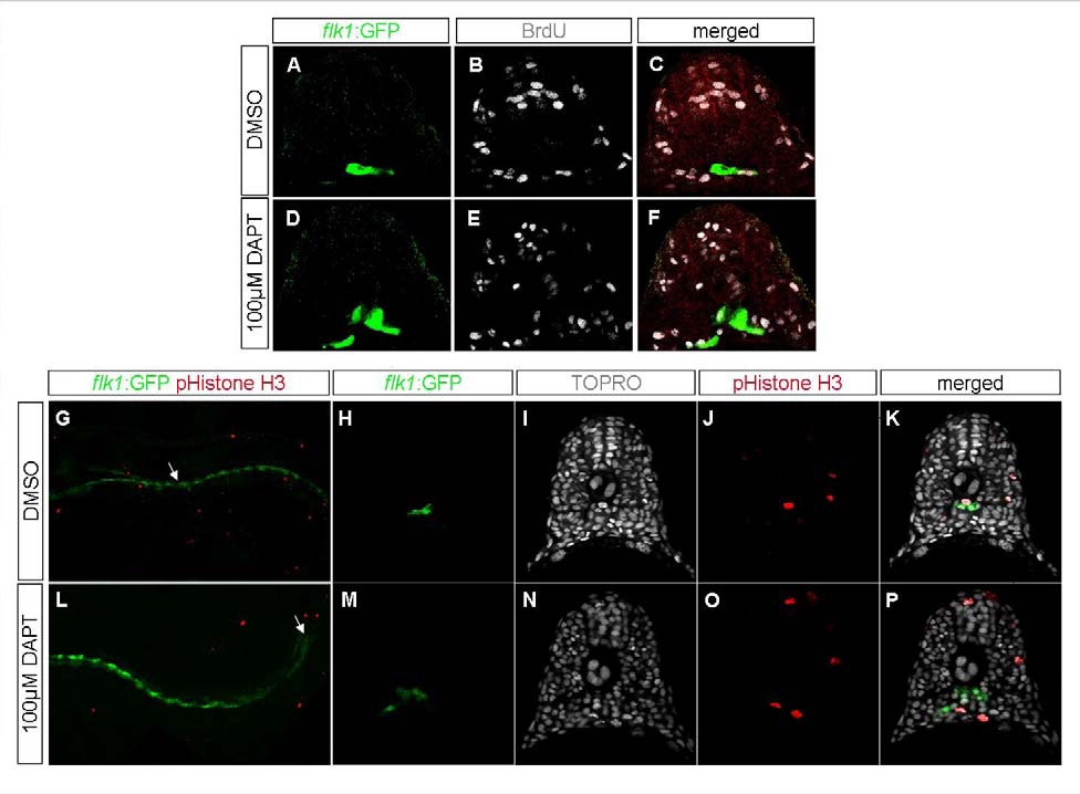
Fig. S6
Cell Proliferation Is Unaffected in Embryos with Reduced Notch Signaling
Transverse sections (A-F, H-K, and M-P) or lateral whole mount view (G and L) of 18 hpf embryos treated with DMSO (A-C and G-K) or DAPT (D-F and L-P), with two-hour pulse of 10mM BrdU starting at 12 hpf (A-F) or stained with anti-phospho-Histone H3 (G-P), visualized for kdrl:GFP (A, D, G, K, L, and K) (green), BrdU (B and E) or TOPRO (I ,K, N, and P) (white), and anti-phospho-Histone H3 (G, J, L, and O) (red). Inhibition of Notch signaling did not increase the number of proliferating endothelial cells.
Acknowledgments
This image is the copyrighted work of the attributed author or publisher, and
ZFIN has permission only to display this image to its users.
Additional permissions should be obtained from the applicable author or publisher of the image.
Full text @ Curr. Biol.