
IMAGE
Fig. S2
- ID
- ZDB-IMAGE-091215-57
- Publication
- Lee et al., 2009 - Notch Signaling Functions as a Cell-Fate Switch between the Endothelial and Hematopoietic Lineages
- All Figures
- Figures for Lee et al., 2009
Image
|
Figure Caption
Fig. S2
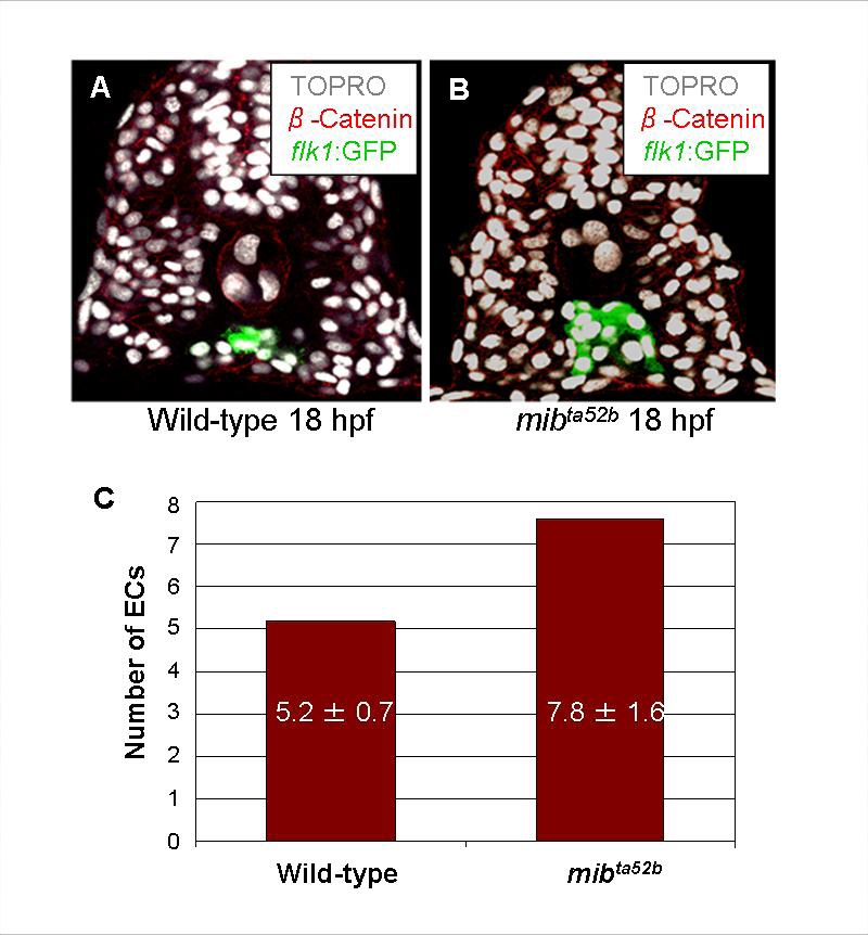
mibta52b Mutant Embryos Display an Increased Number of Endothelial Cells
Transverse sections of 18 hpf embryos, phenotypic wild-type (A) or mibta52b (B), visualized for TOPRO (white), kdrl:GFP (green), and phalloidin (red). (C) The number of endothelial nuclei per focal plane was quantified. Homozygous mibta52b mutant embryos exhibited an increased number of endothelial cells.
Acknowledgments
This image is the copyrighted work of the attributed author or publisher, and
ZFIN has permission only to display this image to its users.
Additional permissions should be obtained from the applicable author or publisher of the image.
Full text @ Curr. Biol.