
IMAGE
Fig. S1
- ID
- ZDB-IMAGE-091215-43
- Publication
- Reischauer et al., 2009 - Lgl2 executes its function as a tumor suppressor by regulating ErbB signaling in the zebrafish epidermis
- All Figures
- Figures for Reischauer et al., 2009
Image
|
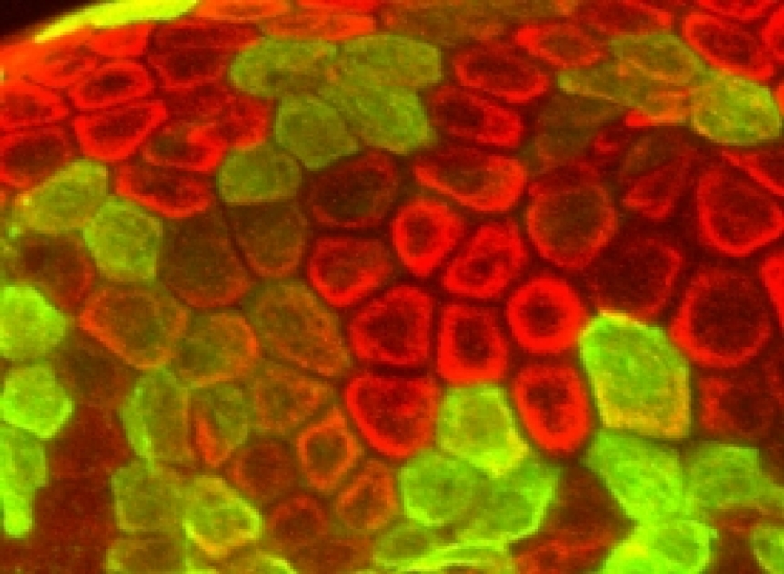
Figure Caption
Fig. S1 Expression of GFP under the ΔNp63 promoter. Antibody staining of 5.5dpf tg(ΔNp63::Gal4,UAS::GFP) zebrafish larvae using anti Cytokeratin (red) and anti GFP antibody (green). The co-labeling of both antibodies reveals activity of the 4.96 kb upstream promoter element of ΔNp63 exclusively in basal epidermal cells in the skin.
Acknowledgments
This image is the copyrighted work of the attributed author or publisher, and
ZFIN has permission only to display this image to its users.
Additional permissions should be obtained from the applicable author or publisher of the image.
Full text @ PLoS Genet.