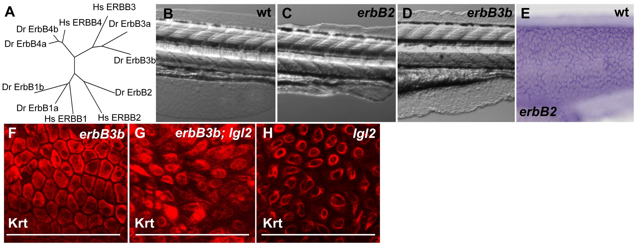
Fig. 5 Phylogenetic analysis of erbB family members and zebrafish mutants of erbB paralogs.
(A) Phylogenetic analysis of erbB family members in the zebrafish genome (zv7) with the human orthologs (Minimum evolution algorithm, 100 replicates). With the exception of erbB2, all other erbB family members are duplicated in zebrafish. (B–D) DIC Images of wild-type (B), erbB2-/- (C) and erbB3b-/- (D) larvae at 132hpf. (E) In-situ hybridization analysis of erbB2 at 48hpf. Keratin staining in erbB3b mutant larva (F) erbB3b/lgl2 double mutant larva (G) and pen/lgl2 mutant larva (H) at 132 hpf. Note that in erbB3B;lgl2 double mutants (G) epidermal cells appear spindle shaped as in pen/lgl2 mutant larvae (H). erbB3b (F) mutations alone do not affect the morphology of basal epidermal cells.

