
IMAGE
Fig. S5
- ID
- ZDB-IMAGE-091201-8
- Publication
- Siekmann et al., 2009 - Chemokine signaling guides regional patterning of the first embryonic artery
- All Figures
- Figures for Siekmann et al., 2009
Image
|
Figure Caption
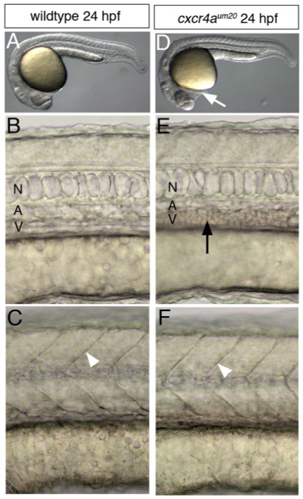
Fig. S5 Phenotypic comparison of wildtype and cxcr4a um20 mutant embryos at 24 hpf. A-C, wildtype, E-F, cxcr4a um20 mutant. Lateral views, anterior to the left. A, D, overview of general morphology. Arrow in B indicates forming edema in cxcr4a um20 mutant embryos. B, E, close-up of axial vasculature. N-notochord, A-dorsal aorta, V-posterior cardinal vein. Black arrow indicates pooled blood cells in the posterior cardinal vein in cxcr4a um20 mutant embryos due to lack of blood flow. C, F, somite boundaries (white arrowheads) form normally in cxcr4a um20 mutant embryos.
Figure Data
Acknowledgments
This image is the copyrighted work of the attributed author or publisher, and
ZFIN has permission only to display this image to its users.
Additional permissions should be obtained from the applicable author or publisher of the image.
Full text @ Genes & Dev.