
Fig. S7
- ID
- ZDB-IMAGE-091201-10
- Genes
- Publication
- Siekmann et al., 2009 - Chemokine signaling guides regional patterning of the first embryonic artery
- All Figures
- Figures for Siekmann et al., 2009
|
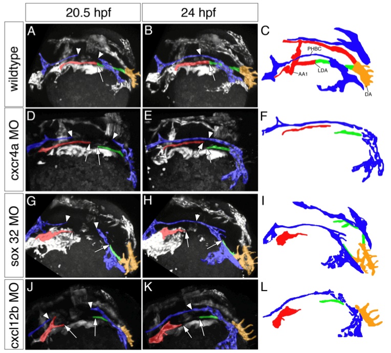
Fig. S7 Stills of 2-photon movies showing defects in lateral dorsal aorta formation. Timepoints are indicated. Aorta is pseudocolored in red, vein is pseudocolored in blue. Arrows mark the positions of the branches of the anterior lateral dorsal aortae (red) and the posterior lateral dorsal aortae (green), respectively. Dorsal aorta cells are pseudocolored in orange. Arrowheads mark the positions of the primordial hindbrain channel branches. C, F, I, L, camera lucida drawings. LDA-lateral dorsal aorta, PHBC-primordial hindbrain channel, DA- dorsal aorta, AA1-aortic arch 1. A-C, wildtype embryo, D-F, embryo injected with cxcr4a morpholino. G-H, sox32 morpholino (MO) injected embryo lacking endoderm. J-K, embryo injected with cxcl12b morpholino. Note the lack of dorsal aorta closure in embryos lacking cxcr4a, cxcl12b or the endoderm (sox32 morpholino injected embryos), while development of the primordial hindbrain channel proceeds normally.