
IMAGE
Fig. 1
- ID
- ZDB-IMAGE-091201-1
- Genes
- Publication
- Siekmann et al., 2009 - Chemokine signaling guides regional patterning of the first embryonic artery
- All Figures
- Figures for Siekmann et al., 2009
Image
|
Figure Caption
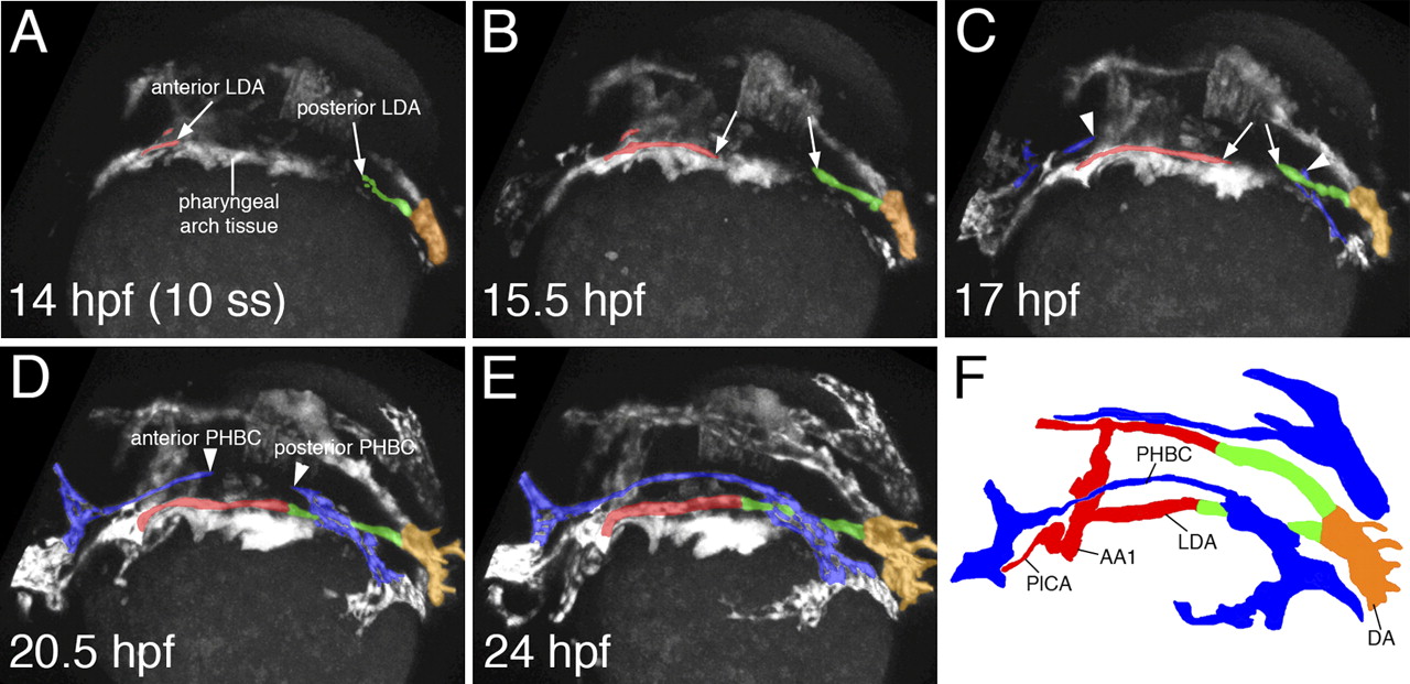
Fig. 1 Two-photon time lapse of LDA formation in live zebrafish embryos. (A–F) Dorsolateral view of LDA formation. Anterior LDA is pseudocolored in red, posterior LDA is shown in green, axial DA is indicated by orange, and the PHBC is shown in blue. Arrows mark migrating cells of the LDA, which forms in a bidirectional manner. Arrowheads indicate similarly migrating PHBC cells. (F) Labeled camera lucida image indicating position of vessels in E. (DA) Dorsal aorta; (LDA) lateral dorsal aorta; (PHBC) primordial hindbrain channel; (PICA) primitive internal carotid artery; (AA1) aortic arch 1.
Figure Data
Acknowledgments
This image is the copyrighted work of the attributed author or publisher, and
ZFIN has permission only to display this image to its users.
Additional permissions should be obtained from the applicable author or publisher of the image.
Full text @ Genes & Dev.