
Fig. S1
- ID
- ZDB-IMAGE-091113-7
- Publication
- Parsons et al., 2009 - Notch-responsive cells initiate the secondary transition in larval zebrafish pancreas
- All Figures
- Figures for Parsons et al., 2009
|
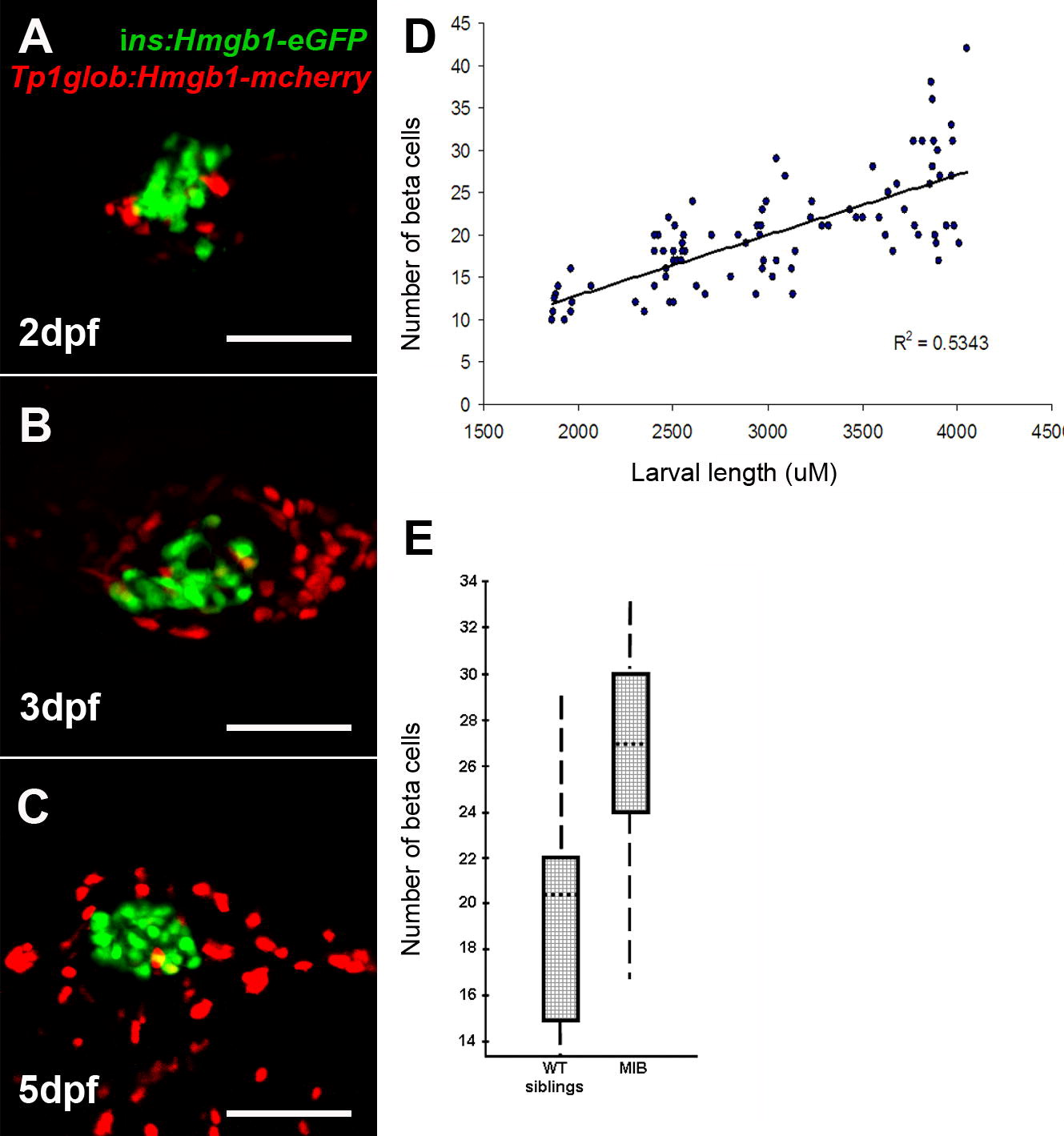
Fig. S1 Quantification of β-cells in the principal islet, and the effects of Notch inhibition. (A–C) islet confocal images from Tg(T2Kins:hmgb1-eGFP; T2KTp1bglob:hmgb1-mCherry) larvae. Pictures generated from multiple optical sections rendered to a single image. Very few PNCs (red nuclei) are associated with the β-cells of the principal islet (green nuclei). No cells were detected that were double positive. (D) Results of measuring larvae and then imaging the head of the pancreas to count β-cell number. Following confocal Z-section analysis through the islet, the number of GFP positive nuclei was plotted against larval length. The graph demonstrates that β-cell number increases with larval size. The same β-cell counting approach was taken with ins:hmgb1-eGFP larvae that were homozygous for the mibta52b (-/-) mutation or phenotypically wildtype (wt) clutch mates (-/+ and +/+). Pancreata from mib and wt larvae were collected at 48 and 72hpf, dissected and imaged by confocal microscopy. β-Cell number for the 2 time points were combined for each group (wt and mib) as there has no significant change in number within each group over this time period (48–72hpf). Comparison of β-cell number in mib (mean = 26.1, n = 10) and wt (20.0, n = 10) is shown in a box plot (E). Boxes represent 50% of the data, with the median marked as a dotted line. The vertical lines represent the whole data range. With mib dependent Notch inhibition, there is a statistically significant, but less than dramatic, effect on β-cell number (ANOVA, p = 0.0123). [Note, when the data from the 2 time points used was not combined, there was still significant increase in β-cells at either 48 hpf (p = 0.048) and 72 hpf (p = 0.033).] This confirms that Notch inhibition, via the mib mutation, leads to increased number of β-cells in the principal islet.
Reprinted from Mechanisms of Development, 126(10), Parsons, M.J., Pisharath, H., Yusuff, S., Moore, J.C., Siekmann, A.F., Lawson, N., and Leach, S.D., Notch-responsive cells initiate the secondary transition in larval zebrafish pancreas, 898-912, Copyright (2009) with permission from Elsevier. Full text @ Mech. Dev.